
CO-CHAIR – Monika Zbroja-Putowska
October 2, 2025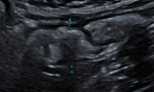
Incidental finding of an appendiceal mucocele [August 2025]
November 21, 2025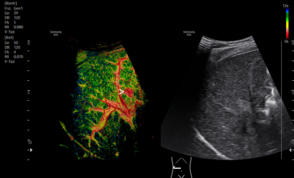
SUBMIT YOUR CASE
EFSUMB invites submission of interesting cases for the website section 'Case of the Month'. All CoM submissions are eligible for selection for free registration at the next Euroson congress. Two cases that receive the most 'likes' in a year will receive free registration for the next EUROSON congress and the third most liked liked case will receive a cash prize of 100 EUR.
Focal nodular hyperplasia revealed with new imaging modalities
Authors:
1. Isabel Wiesinger, Institute for Neuroradiology, Bezirksklinikum Regensburg
2. Ernst Michael Jung, Department of Radiology, Interdisciplinary Center of Ultrasound, University Medical Center Regensburg.
1Clinical History
46 years old female patient with a long history of hormonal replacement therapy presents for evaluation of a unclear liver structure identified during preliminary examination as part of an endocrinological assessment of the liver and abdomen.
The ultrasound examination was performed using a multi-frequency probe (1-7 MHz). The liver was thoroughly examined in B-mode imaging across various standardized ultrasound slices. Additionally, color-coded duplex sonography was used to measure blood flow in the liver veins, the hepatic artery, and the portal vein. Complementary techniques included micro flow imaging in color mode and power Doppler mode. Furthermore, tissue analysis of the liver parenchyma was conducted using shear wave elastography with color coding, with five individual measurements ensuring high image quality, indicated by a minimum of four quality indicators. In addition, a specialized mode for liver fat quantification (QRS) was used to perform analyses with both TAI and TSI sequences.
Contrast-enhanced ultrasound was initially performed in the standard contrast mode, which was then adapted to a higher frequency mode in the high-resolution hepatological setting. Short cine loops were documented for further evaluation.
In B-mode imaging, only minor mixed echogenic structural changes are visible. No clearly defined focal lesions could be identified. Previous examinations, including contrast-enhanced MRI, documented small, highly enhancing liver lesions, the exact nature of which remained undetermined.
The contrast-enhanced ultrasound was performed using bolus technique. After obtaining written consent, 2 ml of ultrasound contrast agent (SonoVue i.v.) was administered as a bolus via a cubital vein.
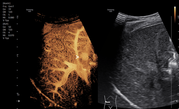
The examination showed rapid contrast enhancement of a focal lesion in the right liver lobe, centrally from the middle to the periphery (Fig.1). No washout phenomenon was observed in the late phase.
The detailed analysis utilized the new high-resolution, color-coded contrast ultrasound technique in Super Resolution mode, MV-CEUS. The false-color analysis with rapid, high contrast from the center to the periphery facilitated the localization of small focal lesions in the right liver lobe (Fig.2).
The patient then mentioned that she stopped taking hormonal replacement therapy. Within one year the lesion remained stable.
The ultrasound examination was performed using a multi-frequency probe (1-7 MHz). The liver was thoroughly examined in B-mode imaging across various standardized ultrasound slices. Additionally, color-coded duplex sonography was used to measure blood flow in the liver veins, the hepatic artery, and the portal vein. Complementary techniques included micro flow imaging in color mode and power Doppler mode. Furthermore, tissue analysis of the liver parenchyma was conducted using shear wave elastography with color coding, with five individual measurements ensuring high image quality, indicated by a minimum of four quality indicators. In addition, a specialized mode for liver fat quantification (QRS) was used to perform analyses with both TAI and TSI sequences.
Contrast-enhanced ultrasound was initially performed in the standard contrast mode, which was then adapted to a higher frequency mode in the high-resolution hepatological setting. Short cine loops were documented for further evaluation.
In B-mode imaging, only minor mixed echogenic structural changes are visible. No clearly defined focal lesions could be identified. Previous examinations, including contrast-enhanced MRI, documented small, highly enhancing liver lesions, the exact nature of which remained undetermined.
The contrast-enhanced ultrasound was performed using bolus technique. After obtaining written consent, 2 ml of ultrasound contrast agent (SonoVue i.v.) was administered as a bolus via a cubital vein.
The examination showed rapid contrast enhancement of a focal lesion in the right liver lobe, centrally from the middle to the periphery (Fig.1). No washout phenomenon was observed in the late phase.
The detailed analysis utilized the new high-resolution, color-coded contrast ultrasound technique in Super Resolution mode, MV-CEUS. The false-color analysis with rapid, high contrast from the center to the periphery facilitated the localization of small focal lesions in the right liver lobe (Fig.2).
The patient then mentioned that she stopped taking hormonal replacement therapy. Within one year the lesion remained stable.
2Teaching Points
Based on the rapid contrast enhancement, the following entities are considered as possible diagnoses for the lesion:
1. High-flow hemangioma with rapid contrast uptake and no washout phenomenon. Typically, this would show high signal intensity in T2-weighted MRI.
2. Adenoma, which tends to show faster contrast enhancement from the periphery to the center, with mixed signals in T1 and T2 MRI images, serving as a differential diagnosis.
3. Focal nodular hyperplasia (FNH), indicated by contrast enhancement from the center to the periphery, resembling a spoke-wheel pattern, supported by the consistency of findings over time.
4. Malignant lesions, such as early-stage hepatocellular carcinoma (HCC), which would likely show irregular vascularization with washout in the late phase.
The newly advanced high-resolution contrast techniques highlight the detailed accuracy of micro flow CEUS in dynamic assessment compared to MRI with liver-specific contrast agents. This allows for a more precise differentiation of benign lesions.
Overall, this case demonstrates the importance of dynamic contrast-enhanced ultrasound, from early arterial phase through portal venous phase and follow-up up to 5-6 minutes in late phase, enabling more accurate differentiation of unclear entities compared to other imaging modalities. The additional examinations performed in this study, such as liver fibrosis assessment and liver fat quantification, also contribute significantly to better characterization of liver lesions.
3References
1. Dietrich CF et al: Guidelines and Good Clinical Practice Recommendations for Contrast-Enhanced Ultrasound (CEUS) in the Liver-Update 2020 WFUMB in Cooperation with EFSUMB, AFSUMB, AIUM, and FLAUS. Ultrasound Med Biol 2020; 46: 2579-2604.
2. Dropco I et al: Color Mapping using Ultrasound System-integrated Perfusion Software for Evaluation of Focal Liver Lesions: A Possible First Step for More Independent Reading. J Gastrointestin Liver Dis. 2023; 32: 479-487.
3. Jung EM et al: Current aspects of multimodal ultrasound liver diagnostics using contrast-enhanced ultrasonography (CEUS), fat evaluation, fibrosis assessment, and perfusion analysis - An update. Clin Hemorheol Microcirc. 2023; 83: 181-193.
