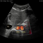
The Double Aorta Artifact: A Benign Trap in Abdominal Sonography [October 2025]
December 8, 2025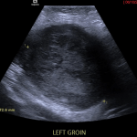
Beyond the visible borders: imaging clues in an infiltrative subcutaneous tumour [December 2025]
December 19, 2025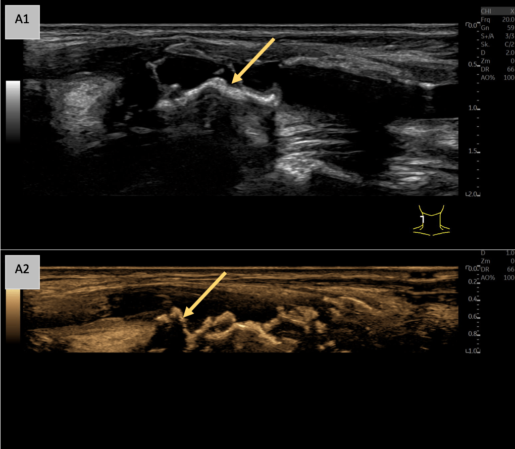
SUBMIT YOUR CASE
EFSUMB invites submission of interesting cases for the website section 'Case of the Month'. All CoM submissions are eligible for selection for free registration at the next Euroson congress. Two cases that receive the most 'likes' in a year will receive free registration for the next EUROSON congress and the third most liked liked case will receive a cash prize of 100 EUR.
Vulnerable Plaque in Focus: A Multimodal Ultrasound Case Study with Triplex, High-Resolution Flow, and 3D Imaging
Authors:
Dr. med. Johannes Matthias Weimer [1], Dr. med. Maximilian Rink [2], Prof. Dr. med. Julian Künzel [2], Prof. Dr. med. Ernst-Michael Jung [3]Affiliation:
[1] Department of Internal Medicine I, University Medical Center of the Johannes Gutenberg University Mainz, Mainz, Germany[2] Department of Otorhinolaryngology, Head and Neck Surgery, University Hospital Regensburg, Regensburg, Germany
[3] Department of Radiology, University Hospital Regensburg, Germany.

A: B-Mode imaging in the sagittal plane (A1) shows a markedly echogenic and inhomogeneous vessel wall of the common carotid artery with a thickened intima-media complex and irregular echogenic wall changes. Notably, multiple echogenic, wall-adherent formations with partial distal acoustic shadowing are observed (yellow arrow), indicative of significant plaque formation. Speckle Reduction Imaging (SRI) and Cross Beam techniques were employed to optimize the B-Mode image (A2), which is illustrated in sepia for enhanced visualization.


D: Visualization of the vessel with the PIUR tUS Infinity v4.1.2 General Imaging App in the transverse plane (D1), reconstructed sagittal plane (D2), and reconstructed coronal plane (D4). The coronal plane particularly highlights the vessel narrowing caused by the plaques (yellow arrow). The additional 3D reconstruction (D3) provides an "external" view of the multiple plaques, illustrating the resulting irregular vessel morphology.
B: B-Mode in sepia and high-resolution flow imaging (B-Flow) along the longitudinal axis of the vessel reveals ulcerated plaque formations with notches and vasa vasorum showing transmural vascular ingrowth into the plaque (red arrow).
C: Triplex, B-Mode, and Duplex imaging with color coding and pulsed wave Doppler reveal significantly increased systolic flow velocities (>200 cm/s) in the CCA, accompanied by aliasing in the region of luminal narrowing (yellow arrow) caused by echogenic plaque formation.
D: Visualization of the vessel with the PIUR tUS Infinity v4.1.2 General Imaging App in the transverse plane (D1), reconstructed sagittal plane (D2), and reconstructed coronal plane (D4). The coronal plane particularly highlights the vessel narrowing caused by the plaques (yellow arrow). The additional 3D reconstruction (D3) provides an "external" view of the multiple plaques, illustrating the resulting irregular vessel morphology.
Triplex ultrasound integrates B-Mode, Color Doppler, and Spectral Doppler to provide comprehensive structural and hemodynamic data, including flow velocity quantification and the detection of microvascular activity (Fig. C). Furthermore, tomographic three-dimensional (3D) ultrasound uniquely enhances understanding of vessel anatomy (Fig. D) and serves as an innovative adjunct imaging modality.
Together, these techniques significantly improve the accuracy of identifying high-risk plaques, aiding in risk stratification and supporting clinical decision-making.
• Comprehensive Hemodynamic Assessment with Triplex Ultrasound: Triplex ultrasound combines B-Mode (structural imaging), Color Doppler (flow visualization), and Spectral Doppler (velocity quantification), providing both anatomical and functional data. This is essential for identifying stenotic lesions, flow velocity changes, and areas of turbulent flow associated with high-risk plaques.
• Enhanced Understanding with 3D Ultrasound: Three-dimensional (3D) ultrasound provides a unique perspective by offering tomographic imaging of vascular anatomy. This modality enables superior spatial visualization of complex plaque morphology and vessel wall structures, aiding in identifying subtle irregularities and refining risk assessment.
2. Hofmann AG, Mlekusch I, Wickenhauser G et al. Clinical Applications of B-Flow Ultrasound: A Scoping Review of the Literature. Diagnostics (Basel) 2023; 13. doi:10.3390/diagnostics13030397
3. Jung EM, Clevert DA, Rupp N. [B-flow and color-coded B-flow in sonographic diagnosis of filiform stenosis of the internal carotid artery]. Rofo 2003; 175: 1251-1258. doi:10.1055/s-2003-41936