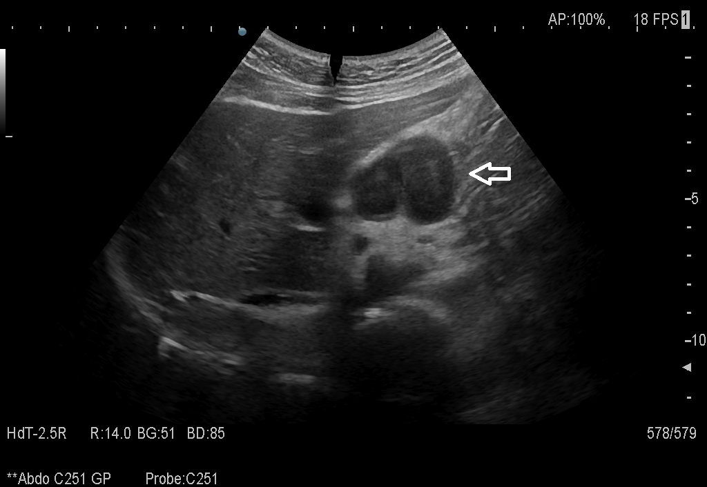
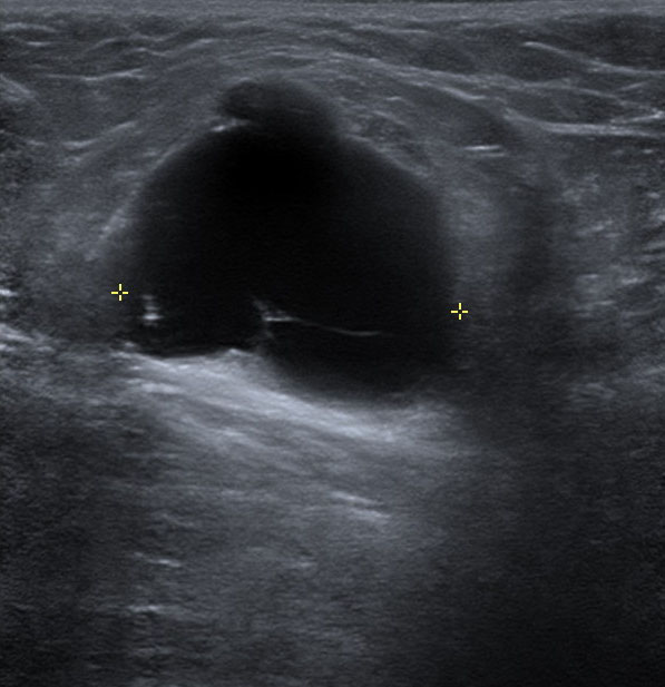
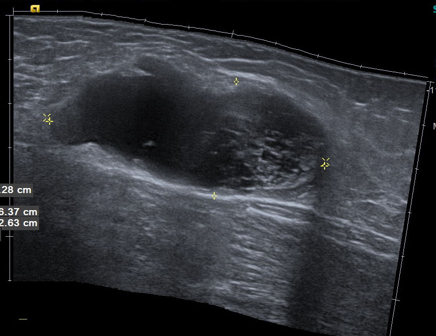
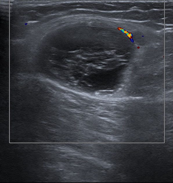
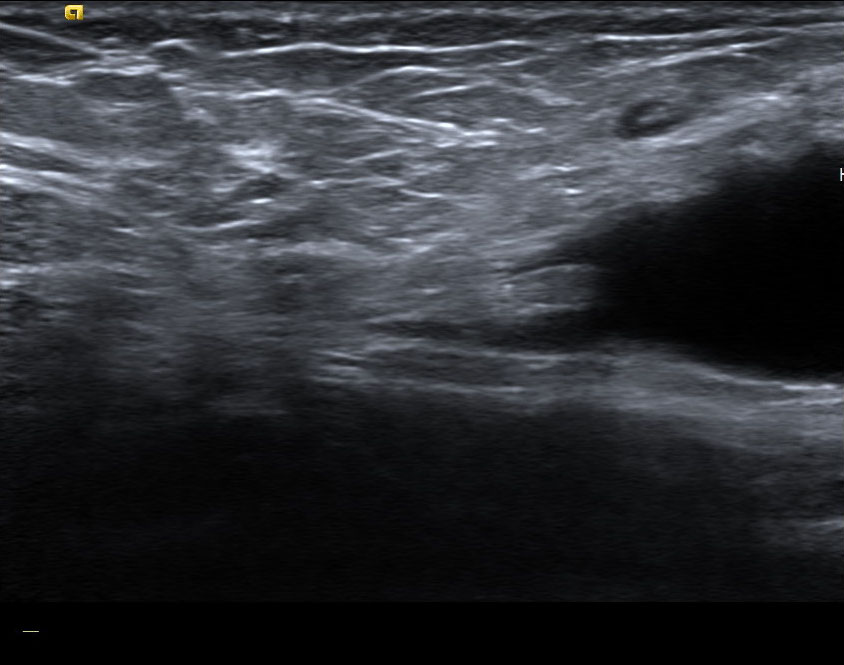
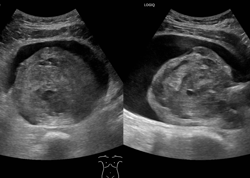
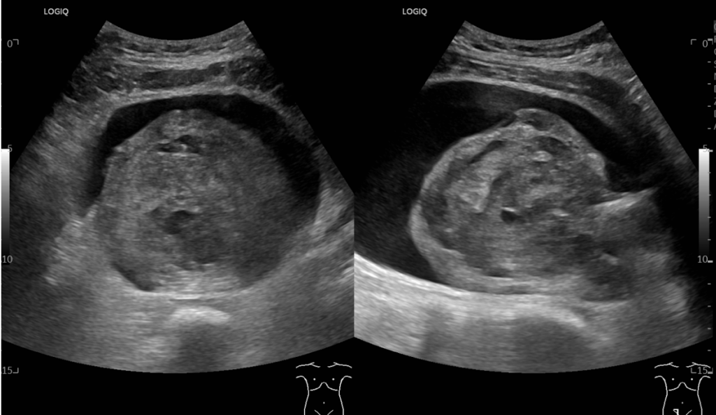
Student Image Challenge 89April 30, 2021WFUMB / EFSUMB Students webinar seriesMay 5, 2021 Student Image Challenge 90 Image 01 Image 02 Image 03 Image 04 Image 05 Student Image Challenge #90 1 / 1 Student Image Challenge #90 A 40-year-old patient with a good general state, presented for right inguinal swelling, moderately painful, appearing after repeated physical efforts, slowly evolving. Identify the indicated mass. What would be your diagnosis? Groin hematoma Round ligament rupture Herniated fluid accumulation Incarcerated inguinal hernia with small bowel loop Incorrect ....Please see the correct answer highlighted Correct: Round ligament rupture Image 1 & 2. High frequency probe revealed an inguinal cystic structure, irregularly delimited, well contoured, not encapsulated, inhomogeneous by the presence of fibrillar structures located at the lower pole. Image 3. No cough impulse visible. The echoes did not mobilize in real time. It was not related to the inguinal vascular elements and there were no visible vessels on Doppler colour. Image 4. The analysis of the upper pole of the lesion reveals the amputated fibrillar structure which is visible in the rest of the groin region. Image 5. The panoramic image shows the relationship between fluid accumulation and the fibrillar structure of the upper pole. Your score isThe average score is 0% LinkedIn Facebook VKontakte 0% Restart quiz Case courtesy of Dana Nedelcu MD EFSUMBAdmin Related postsStudent Image Challenge 114Read more Comments are closed.
