EFSUMB Young Investigators’ Awards 2020
June 12, 2020
UNITED KINGDOM – BMUS
July 7, 2020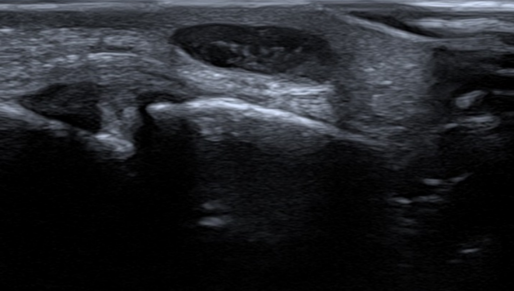
Is it cystic or solid?
AUTHORS:
Dr Sabrina Memarian, Radiology registrar
Dr Mohammed Riyaz, Radiology registrar
Professor Adrian Lim, Professor and Consultant Radiologist
Imaging Department, Imperial College Healthcare NHS Trust, London
Dr Sabrina Memarian, Radiology registrar
Dr Mohammed Riyaz, Radiology registrar
Professor Adrian Lim, Professor and Consultant Radiologist
Imaging Department, Imperial College Healthcare NHS Trust, London
![Is it cystic or solid? </br> [Jun/Jul 2020]](http://s834315022.websitehome.co.uk/wp-content/uploads/2020/11/june2020-fig01.png)
1Clinical history
The patient presented to the plastic surgery clinic on 02/12/2019 due to a lump on the inner dorsal aspect of the left middle finger. He initially had a radiograph of the left middle and index fingers which was performed on 02/12/2019 and was normal. Subsequently, a month later on 06/01/2020 an ultrasound of the left middle finger was performed to ascertain the nature of this persisting lump.
2Image Findings
The palpable nodule along the radial aspect adjacent to the PIPJ of the left middle finger corresponded to an oval shaped subcutaneous lesion which contained internal echoes and on first inspection appears solid (fig.1). However, on turning on color doppler, there was clear movement of the internal echoes within this lesion suggesting that it was cystic with proteinaceous material rather than being solid. This changed the differential diagnosis significantly. In addition, there was no definite connection with the PIPJ but appeared to abut the tendon sheath.
3Diagnosis
The appearances, particularly with the Doppler findings, confirmed that this was a ganglion cyst with proteinaceous material.
4Discussion
Ultrasound is usually the first modality of choice to evaluate a superficial palpable lump and can readily distinguish solid from cystic lesions with good accuracy. However, there are times where a lesion can look solid and the utility of other ultrasound modes can be helpful in achieving the correct diagnosis.
A Ganglionic cyst on ultrasound typically appears as a well-defined anechoic to hypoechoic fluid filled lesion, with an ovoid or irregular shape, and a stalk extending towards the joint, which is not compressible.
Ganglionic cysts with thick proteinaceous material however can mimic the ultrasound appearance of solid masses and be mistaken for tumors. In this case, Doppler was crucial for the diagnosis where at first glance, the lesion appears solid on B mode. However, the acoustic streaming effect of colour/power Doppler confirmed the cystic nature of this lesion, which cannot be induced in solid lesions. Acoustic streaming is the utilization of Doppler technique, whereby absorption of high amplitude acoustic oscillations results in the continuous flow of fluid. This explains why we can observe the movement of the proteinaceous material within the ganglion cyst (fig. 2). This simple, yet very effective application of Doppler can help avoid a further MRI or invasive biopsy.
5Background
Ganglia are benign, mucin-filled cystic lesions associated with joints or tendon sheaths. Patients present with a palpable lump or pain, most commonly arising in the wrist or hand. Ganglion cysts are known to resolve without treatment; however aspiration or surgical excision may be indicated if the ganglion is causing pain, stiffness, or nerve compression.
6Clinical Perspective
Clinical and ultrasonographic findings of the lesion navigate the potential for further imaging and/or intervention.
7Therapy Planning
Conservative management unless there is a clinical need for surgical excision where the lesion obstructs movement and is painful.
8Outcome
The patient was reassured and discharged home with a provisor, that should the lesion grow, cause pain or get infected, then medical advice should be sought.
9Prognosis
Most ganglion cysts are self-limiting and resolve without treatment. In a small proportion however, these cysts can recur despite excision or aspiration.
10Teaching Points
The Doppler effect of acoustic streaming was extremely useful in confirming the cystic nature of this lesion, which on first inspection appeared solid. This significantly altered the diagnosis and further management of the patient.
11References
1. Bianchi S, Abdelwahab IF, Zwass A, Giacomello P. Ultrasonographic evaluation of wrist ganglia. Skeletal Radiol 1994; 23:201–203.
2. De Flaviis L, Nessi R, Del Bo P, Calori G, Balconi G. High-resolution ultrasonography of wrist ganglia. J Clin Ultrasound 1987; 15:17–22.
3. Paivansalo M, Jalovaara P. Ultrasound findings of ganglions of the wrist. Eur J Radiol 1991; 13:178–180.
4. Aditi, G., Vilas, D.K., & Amarjeet, D.S. (2014). Acoustic streaming ( Non-vascular application of colour Doppler ) in differentiating complicated cyst and solid lesion in breast – a case study and review of literature.
