Basic Obstetric Scanning
January 25, 2019CEUS for guidance and monitoring of interventions
March 17, 2019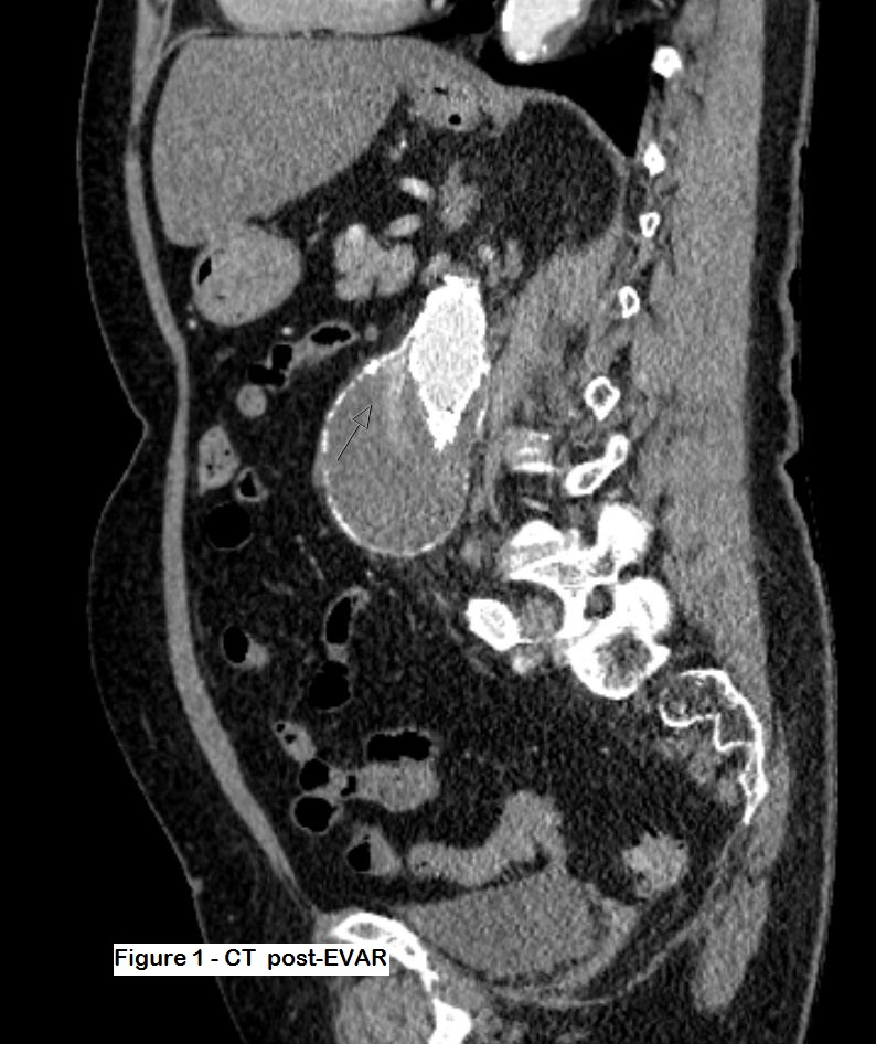
Post-EVAR control with CEUS and fusion-ultrasound
Authors:
Dr Sara Sehlstedt, Radiology Dept, Östersunds Sjukhus
Dr Sara Sehlstedt, Radiology Dept, Östersunds Sjukhus
![Post-EVAR control with CEUS and fusion-ultrasound </br> [Feb 2019]](http://s834315022.websitehome.co.uk/wp-content/uploads/2020/11/PostEVAR1-version-fig01.jpg)
![Post-EVAR control with CEUS and fusion-ultrasound </br> [Feb 2019]](http://s834315022.websitehome.co.uk/wp-content/uploads/2020/11/PostEVAR1-version-fig02.jpg)
![Post-EVAR control with CEUS and fusion-ultrasound </br> [Feb 2019]](http://s834315022.websitehome.co.uk/wp-content/uploads/2020/11/PostEVAR1-version-fig03.jpg)
![Post-EVAR control with CEUS and fusion-ultrasound </br> [Feb 2019]](http://s834315022.websitehome.co.uk/wp-content/uploads/2020/11/PostEVAR1-version-fig04.jpg)
![Post-EVAR control with CEUS and fusion-ultrasound </br> [Feb 2019]](http://s834315022.websitehome.co.uk/wp-content/uploads/2020/11/PostEVAR1-version-fig05.jpg)
![Post-EVAR control with CEUS and fusion-ultrasound </br> [Feb 2019]](http://s834315022.websitehome.co.uk/wp-content/uploads/2020/11/PostEVAR1-version-fig06.jpg)
![Post-EVAR control with CEUS and fusion-ultrasound </br> [Feb 2019]](http://s834315022.websitehome.co.uk/wp-content/uploads/2020/11/PostEVAR1-version-fig07.jpg)
![Post-EVAR control with CEUS and fusion-ultrasound </br> [Feb 2019]](http://s834315022.websitehome.co.uk/wp-content/uploads/2020/11/PostEVAR1-version-fig08.jpg)
![Post-EVAR control with CEUS and fusion-ultrasound </br> [Feb 2019]](http://s834315022.websitehome.co.uk/wp-content/uploads/2020/11/PostEVAR1-version-fig09.jpg)
![Post-EVAR control with CEUS and fusion-ultrasound </br> [Feb 2019]](http://s834315022.websitehome.co.uk/wp-content/uploads/2020/11/PostEVAR1-version-fig10.jpg)
![Post-EVAR control with CEUS and fusion-ultrasound </br> [Feb 2019]](http://s834315022.websitehome.co.uk/wp-content/uploads/2020/11/PostEVAR1-version-fig11.jpg)
1Clinical History
The patient is a well preserved lady of 90 years, with a medical history of high blood pressure and well controlled diabetes. Due to an abdominal aortic aneurysm (AAA), she had successful intervention with EVAR.
No endoleak was noted during the intervention, but at the first CT follow-up a month later, there was a suspected endoleak. It was however difficult to ascertain whether this was a type 1 or type 2 leak. The patient also had impaired renal function after the procedure, so further use of iodinated contrast was considered to be inappropriate. Therefore, a contrast enhanced ultrasound study (CEUS) was performed instead to see if it could determine the type of endoleak.
No endoleak was noted during the intervention, but at the first CT follow-up a month later, there was a suspected endoleak. It was however difficult to ascertain whether this was a type 1 or type 2 leak. The patient also had impaired renal function after the procedure, so further use of iodinated contrast was considered to be inappropriate. Therefore, a contrast enhanced ultrasound study (CEUS) was performed instead to see if it could determine the type of endoleak.
2Imaging
Figures 1 and 2: Post-op CT which shows some contrast within the aneurysm sac but it is difficult to ascertain the type of leak. The patient was thus referred for an ultrasound study.
Figure 3: Doppler ultrasound shows flow within the aneurysm sac
Figure 4: Ultrasound fusion with the postop CT to better delineate the anatomy. This would also provide more anatomical information to the vascular surgeon and interventional radiologist for future intervention. The Doppler shows very clear colour flow signal within the sac, where it is apparent that it is a type 2 leak arising from the lumbar arteries.
Figure 5: CEUS with fusion ultrasound. A total of three separate injections of Sonovue 1.5 ml intravenously were undertaken. The first injection, longitudinal scan of the aorta, shows a small endoleak type 1A at the top of the stent. Typical for this type of leak is that it occurs simultaneously as the first flow of contrast appears within the aorta/stent.
Figures 6 & 7: Two further injections of contrast with images focused lower down in the stent graft, where there was Doppler flow in the stent. Note the time difference where the first passage of contrast through the graft (figure 6) occurs at 10 seconds after injection, but the endoleakage only appears after 19 seconds. There is thus a delay of nine seconds which is a strong indicator that this is a type 2 endoleak. (time is shown at the bottom of the picture, A = seconds after injection).
Figure 8: US/CT fusion also provides a better understanding of the anatomy, which can be very challenging at times. In this case, the type 2 endoleak extends a long distance throughout the sac.
Figure 9: Delayed images shows “pooling” of US contrast (Sonovue) in the ventral portion of the aneurysm sac, probably due to both endoleakages.
3Discussion
Endoleakages are a common complication after EVAR, and many of these patients also suffer from renal failure due to the underlying cardiovascular disease. CEUS is a very useful tool to help characterise the type of endoleak, which is very important to aid the surgical management of these complications.
In this case, the type 1 leakage was very small and the type 2 leak significantly larger. Owing to the advanced age and comorbidity of our patient, this has been treated conservatively and followed regularly with CT (unenhanced) to ensure that the aneurysm sac was not growing in size (implying a low pressure endoleak). The common practice otherwise for type 1 endoleaks are that they should be repaired as they are considered “high-pressure” leakages and thus a risk for rupture of the aneurysm sac. Type 2 leaks are considered to be ”low-pressure” and therefore usually do not need further invervention unless the aneurysm-sac is enlarging.
In this case, the type 1 leakage was very small and the type 2 leak significantly larger. Owing to the advanced age and comorbidity of our patient, this has been treated conservatively and followed regularly with CT (unenhanced) to ensure that the aneurysm sac was not growing in size (implying a low pressure endoleak). The common practice otherwise for type 1 endoleaks are that they should be repaired as they are considered “high-pressure” leakages and thus a risk for rupture of the aneurysm sac. Type 2 leaks are considered to be ”low-pressure” and therefore usually do not need further invervention unless the aneurysm-sac is enlarging.
4Teaching Points
CEUS is an extremely helpful tool to detect endoleakages post EVAR, especially in patients with renal impairment. It also provides valuable added information to determine the type of leakage. The time difference in contrast agent arriving in the aorta and the different leakages is vital to understand the flow within the sac, and CEUS provides excellent real time visualisation of this important information.
Ultrasound images, and in particular CEUS-images, can sometimes be difficult for surgeons and other colleagues to interpret. US /CT fusion-can thus be a useful aid to allow surgeons and other radiologists to better understand the anatomy and type of endoleak which has been demonstrated.
Ultrasound images, and in particular CEUS-images, can sometimes be difficult for surgeons and other colleagues to interpret. US /CT fusion-can thus be a useful aid to allow surgeons and other radiologists to better understand the anatomy and type of endoleak which has been demonstrated.
5References
Figure 10+11 includes diagrams from Raediopedia on the classification on different types of endoleaks. Excellent article on aortic aneurysm and endoleaks post-EVAR:
https://radiopaedia.org/articles/endoleak?lang=us
