Elasto Small Parts (1)
March 25, 2019Elasto Small Parts (2)
April 25, 2019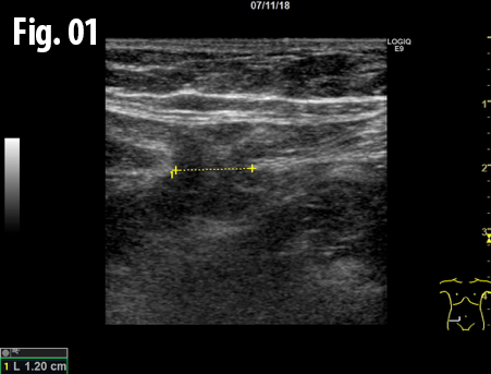
Spigelian hernia
AUTHORS: Meidahl S, Ewertsen C,
Department of Radiology, Rigshospitalet - Copenhagen University Hospital, Denmark
Department of Radiology, Rigshospitalet - Copenhagen University Hospital, Denmark
![Spigelian hernia </br> [Apr 2019]](http://s834315022.websitehome.co.uk/wp-content/uploads/2020/11/cotm_april2019-fig01.png)
![Spigelian hernia </br> [Apr 2019]](http://s834315022.websitehome.co.uk/wp-content/uploads/2020/11/cotm_april2019-fig02.png)
![Spigelian hernia </br> [Apr 2019]](http://s834315022.websitehome.co.uk/wp-content/uploads/2020/11/cotm_april2019-fig03.png)
![Spigelian hernia </br> [Apr 2019]](http://s834315022.websitehome.co.uk/wp-content/uploads/2020/11/cotm_april2019-fig04.png)
1Clinical History
A 55-year-old female presented to our clinic with a history of intermittent pain in the lower right quadrant of the abdomen and also an impression of a lump. These symptoms had been intermittent for approximately six years. The pain was described as a feeling of heaviness and fullness including a dull pain radiating to the flank with intermittent duration – ranging from weekly to monthly. The symptoms and lump seemed to appear after long periods of standing or walking and also after meals.
Clinical examination revealed no palpable lump in the region of interest and an appendicectomy scar was noted medially. The patient had not sought medical advice previously.
Clinical examination revealed no palpable lump in the region of interest and an appendicectomy scar was noted medially. The patient had not sought medical advice previously.
2Imaging Findings
An ultrasound examination of the abdomen was performed using a GE Logiq E9 system with a linear array transducer and centre frequency of 9 MHz. The patient was examined in the supine and standing position including the Valsalva manoeuvre. The examination revealed an incarcerated hernia between the rectus abdominis and transversus abdominis muscles at the level of the anterior superior iliac spine. The dimension of hernial orifice was 1.3 x 0.7 cm and contained preperitoneal fat without any bowel loops. There was no change in size or content of the hernia on standing or Valsalva, and no signs of strangulation.
3Diagnosis
The findings were consistent with a lateral ventral hernia, or Spigelian hernia, containing preperitoneal fatty tissue.
4Discussion
Background: The Spigelian fascia is located lateral to the rectus abdominis muscle along the semilunar line. Its definition is somewhat vague but it is often used synonymously with the aponeurosis of the transversus abdominis muscle1–3. Hernias through this fascia are called Spigelian hernias. They are very rare and constitute approximately 1% of all abdominal hernias1. They are slightly more common in female than in male patients and usually in patients above 40 years1–3.
Eighty-five to ninety percent of Spigelian hernias occur in a transverse band 0-6 cm cranially to the plane between the anterior, superior iliac spines, also called Spigelian hernia belt, which is where the fascia is widest2,3. It has been hypothesized that Spigelian hernias may be related to previous surgery or distention of the abdominal wall due to obesity or pregnancy, or as a complication of peritoneal dialysis3.
Patients can present with a visible and palpable lump in the area, in which case the diagnosis is apparent, but this is rare.
In some cases, the hernia only penetrates the transverse abdominal aponeurosis, but since this is tightly bound to the internal oblique muscle, the hernia usually penetrates both3. Because the external oblique aponeurosis is thick and not bound as tightly to the others, it is usually not penetrated, and the hernia tends to slide between the oblique muscles, and rarely reaches the subcutaneous tissue3.
In addition to the hernias often being obscured by at least one aponeurotic layer, they tend to be small, and they can therefore be very difficult to see and feel3. Symptoms are generally vague, intermittent and unspecific, and although pain is the most common symptom, there is no typical kind of pain associated with Spigelian hernias1–3. The symptoms can mimic other conditions such as appendicitis, abscesses, peptic ulcers, or cholecystitis2. Diagnosis from clinical presentation is often problematic, but can be made by ultrasound or CT with high accuracy1,2. CT shows the anatomy in detail, and is more sensitive but is also more expensive, and a source of radiation. Ultrasound, though slightly less sensitive, is more dynamic as the patient can be examined in supine and upright position and during Valsalva manuevre. In addition, the radiologist can be helped by the patient, who may be able to directly point out the localization of their symptoms during the examination. If doubt persists after radiological examinations then a diagnostic laparoscopy is usually performed2.
Clinical perspective: Spigelian hernias are rare and difficult to diagnose, but diagnosis is important due to the high risk of strangulation owing to their usually small hernial ring.
Therapy planning: The hernias are usually treated surgically, either by open surgery or laparoscopy, with insertion of a mesh intra-, or extraperitoneally to close the hernial ring.
Outcome: The desired outcome is to relieve symptoms and avoid strangulation and recurrence.
Prognosis: There is a risk of recurrence after surgery, but this is generally small1–3.
5Figure legends
Figure 1: The hernia containing preperitoneal fat, axial view obtained with the patient standing. The marker indicates the hernia, the blue arrow the transversus abdominis muscle and the gray arrow indicates the rectus abdominis muscle.
Figure 2: The hernia containing preperitoneal fat, sagittal view, patient standing. The marker indicates the hernia.
Figure 3 and 4: Comparison between right (left side) and left (right side) side, axial view obtained with the patient standing. The right and left rectus muscle (gray arrows) is located centrally in the image. The black arrow and marker indicate the hernia and the blue arrows indicate the transversus abdominis muscles. The yellow arrow indicates the intact fascia on the left side.
6Teaching Points
Although these hernias are very rare it is important to remember them when dealing with a patient with vague symptoms and recurring lower abdominal pain.
7References
1. https://radiopaedia.org/articles/spigelian-hernia-1
2. Mittal T, Kumar V, Khullar R et al. Diagnosis and management of Spigelian hernia: A review of literature and our experience. J Minim Access Surg. 2008;4:95-8.
3. Spangen L. Spigelian hernia. World J Surg. 1989;13:573-80.
2. Mittal T, Kumar V, Khullar R et al. Diagnosis and management of Spigelian hernia: A review of literature and our experience. J Minim Access Surg. 2008;4:95-8.
3. Spangen L. Spigelian hernia. World J Surg. 1989;13:573-80.
