
Past Meetings of ECMUS Safety Committee
October 27, 2020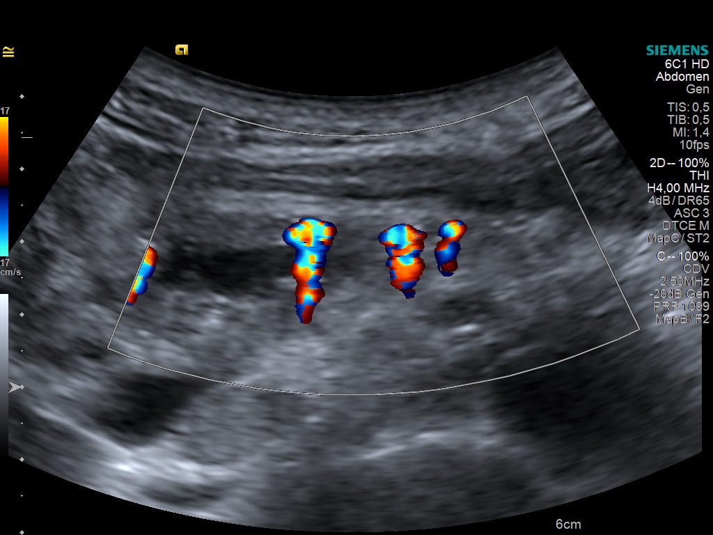
Student Image Challenge 72
November 3, 2020
-
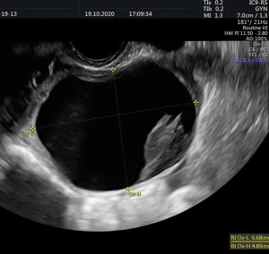
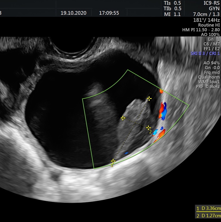
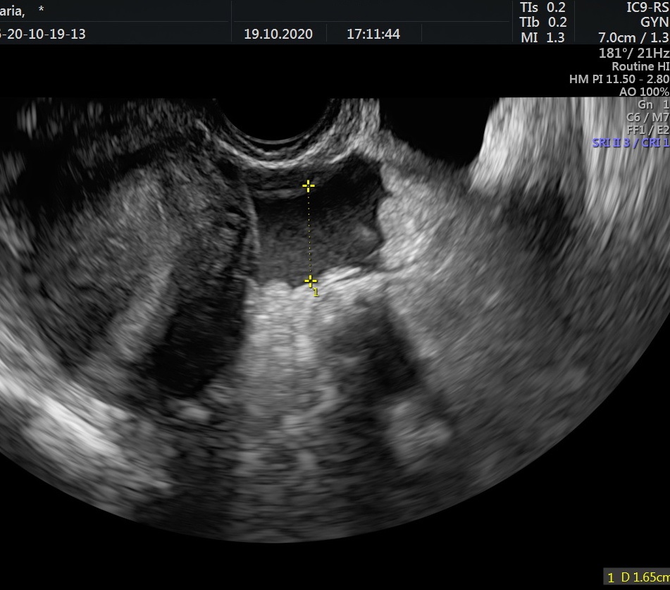
- Fig 01 – transvaginal ultrasound
-

- Fig 02 – transvaginal ultrasound
-
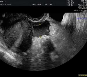
- Fig 03 – transvaginal ultrasound
-
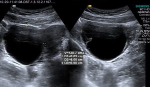
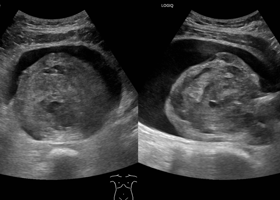
- Fig 04 – transabdominal ultrasound

-
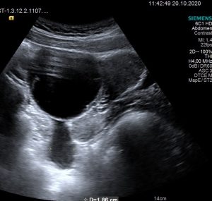
- Fig 05 – transabdominal ultrasound

-
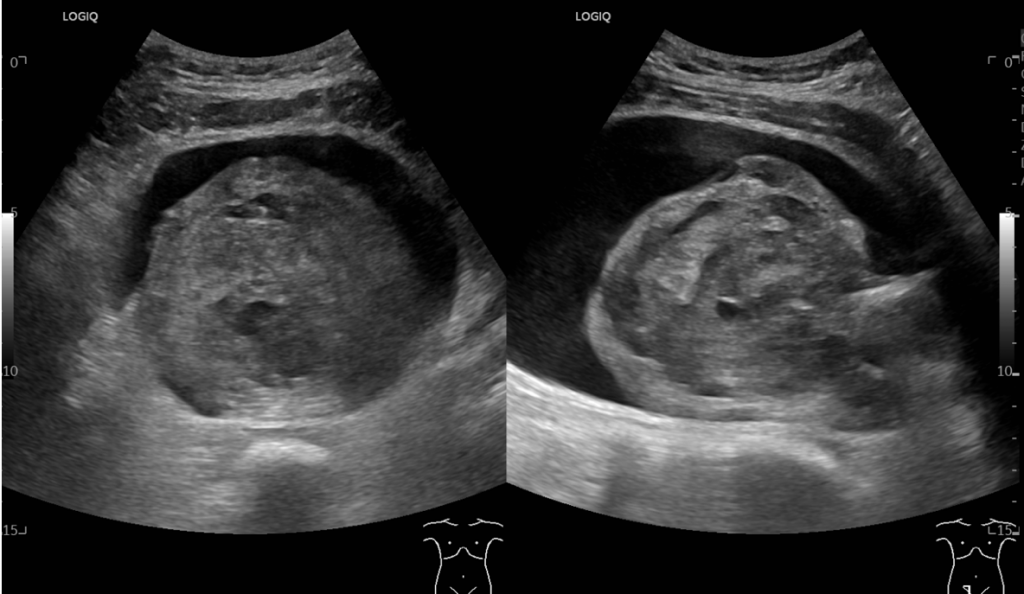
- Fig 06 – transabdominal ultrasound

-
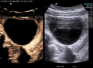
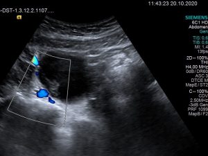
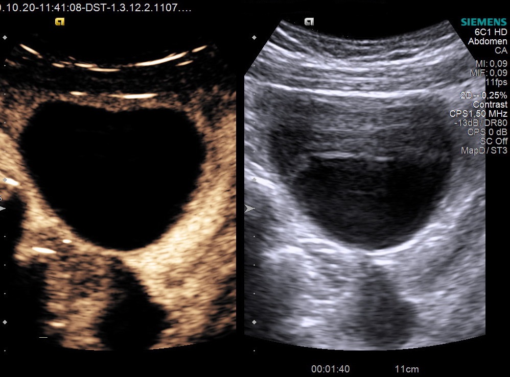
- Fig 07 – contrast enhanced ultrasound (CEUS)
Case courtesy of Dana Nedelcu MD