CEUS for Monitoring of Interventions
May 12, 2020New applications of ultrasound in dermatology
June 2, 2020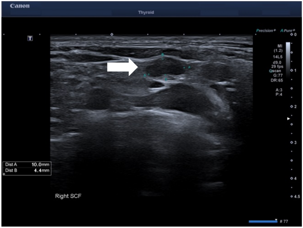
Use of Superb Microvascular Imaging monochrome in evaluating lymph node microvasculature in a young patient with breast cancer
AUTHORS:
Andrew Logan, Adrian KP Lim Imaging Department, Imperial College London and Healthcare NHS Trust, UK
Andrew Logan, Adrian KP Lim Imaging Department, Imperial College London and Healthcare NHS Trust, UK
![Use of Superb Microvascular Imaging monochrome in evaluating lymph node microvasculature in a young patient with breast cancer </br> [May 2020]](http://s834315022.websitehome.co.uk/wp-content/uploads/2020/11/may2020-fig1.png)
![Use of Superb Microvascular Imaging monochrome in evaluating lymph node microvasculature in a young patient with breast cancer </br> [May 2020]](http://s834315022.websitehome.co.uk/wp-content/uploads/2020/11/may2020-fig2.png)
![Use of Superb Microvascular Imaging monochrome in evaluating lymph node microvasculature in a young patient with breast cancer </br> [May 2020]](http://s834315022.websitehome.co.uk/wp-content/uploads/2020/11/may2020-fig03.png)
![Use of Superb Microvascular Imaging monochrome in evaluating lymph node microvasculature in a young patient with breast cancer </br> [May 2020]](http://s834315022.websitehome.co.uk/wp-content/uploads/2020/11/may2020-fig04.png)
1Clinical history
A 29 year old female presented to the same day breast lump clinic with a self-detected tender right breast lump and palpable axillary lymph nodes. She was previously fit and well with no significant family history.
Ultrasound showed a corresponding 20mm mass extending posteriorly into chest wall with enlarged nodes in the axilla. A core biopsy showed grade 3 invasive ductal carcinoma (IDC) T3 P3 M2. Axillary FNA was positive for metastatic disease. Patient was then commenced on chemotherapy.
Owing to the aggressive cancer in a young patient, a staging PET/CT study was performed which showed a small but metabolically active lymph node in the right supraclavicular fossa. An US of the supraclavicular fossa revealed several small volume nodes. The most prominent 10 x 4 mm node had a normal architecture but with only mild cortical thickening. Power Doppler imaging (PDI) and color Doppler imaging (CDI) showed feeding vessels but otherwise no suspicious features. However, on using Superb Microvascular Imaging monochrome (SMI), this revealed abnormal peripheral vascularity which was best depicted in the monochrome mode. This feature made the node suspicious for malignancy and corresponded to the FDG avid node on the PET study. Subsequent fine needle aspirate of the right supraclavicular node confirmed metastatic disease.
Patient went on to have right skin sparing mastectomy and right level III axillary node clearance as well right level 4/5 neck dissection.
2Final Diagnosis
Right breast invasive ductal carcinoma with supraclavicular node spread.
3Imaging Findings
Longitudinal ultrasound of right supraclavicular fossa showed a small 10 x 4 mm lymph node in the right supraclavicular fossa (SCF) with mild cortical thickening. SMI revealed abnormal peripheral vascularity which made it suspicious for metastatic infiltration and most likely represented the metabolically active SCF node on the recent PET imaging.
4Diagnosis
Right breast invasive ductal carcinoma with supraclavicular node metastasis T3N1M0.
5Discussion
Background: There are a growing number of clinical applications using SMI to evaluate diseases closely associated with angiogenesis, including evaluating lesions in liver, breast, thyroid and skeletal muscle. SMI separates flow from overlaying tissue motion preserving subtle low-flow components which gives improved detail and definition compared to PDI and CDI. The SMI monochrome mode removes anatomical background information and reveals fine vasculature with high sensitivity. SMI has recently been shown to be more effective in assessing breast and thyroid lesions than PDI and CDI. It may offer a safe and low-cost alternative to Contrast Enhanced Ultrasound (CEUS).
Clinical perspective: PDI and CDI ultrasound showed relatively normal vascularity in the SCF Lymph node, however, SMI, particularly monochrome mode, outlined clearly the abnormal peripheral vascularity which allowed FNA of the correct “small” node which was FDG avid on the PET study . A recent study has shown SMI yields more detailed information about nodal vessel when compared to PDUS and can be useful in differentiating between malignant and benign lymph nodes.
Therapy planning: Chemotherapy was initially started on diagnosis of invasive ductal breast carcinoma, however, this was stopped when a positive supraclavicular lymph node was identified. Patient agreed to proceed with mastectomy, axillary node clearance, as well as neck dissection.
Outcome: Patient went on to have right skin sparing mastectomy, right level III axillary node clearance and right level 4/5 neck dissection. Histology showed multiple small foci of grade 2 invasive ductal carcinoma and lymph nodes free of tumour. The level 4&5 nodes, axillary apical tissue, infraclavicular and internal mammary lymph nodes were free of tumour.
Prognosis: Stage 3a based on T3N1M0. 72% 5-year survival for stage 3.
6Teaching Points
SMI monochrome mode is more sensitive in assessing lymph node peripheral vasculature compared to PDI and CDI and may be useful in assessing lymph nodes in patients with known breast cancer to differentiate between malignant and benign lymph nodes. In this case, in helped determine the correct node to sample, confirming disease infiltration and thus altered patient management.
7References
1. Jiang ZZ, Huang YH, Shen HL, Liu XT. Clinical Applications of Superb Microvascular Imaging in the Liver, Breast, Thyroid, Skeletal Muscle, and Carotid Plaques. J Ultrasound Med. 2019 Nov;38(11):2811-2820. doi: 10.1002/jum.15008. Epub 2019 Apr 5.
2. Park AY, Seo BK. Up-to-date Doppler techniques for breast tumor vascularity: superb microvascular imaging and contrast-enhanced ultrasound. Ultrasonography. 2018 Apr;37(2):98-106. doi: 10.14366/usg.17043. Epub 2017 Aug 19.
3. Lu R, Meng Y, Zhang Y, Zhao W, Wang X, Jin M, Guo R. Superb microvascular imaging (SMI) compared with conventional ultrasound for evaluating thyroid nodules. BMC Med Imaging. 2017 Dec 28;17(1):65. doi: 10.1186/s12880-017-0241-5.
4. Bakdik S, Arslan S, Oncu F, Durmaz MS, Altunkeser A, Eryilmaz MA, Unlu Y. Effectiveness of Superb Microvascular Imaging for the differentiation of intraductal breast lesions. Med Ultrason. 2018 Aug 30;20(3):306-312. doi: 10.11152/mu-1433.
5. Sim JK, Lee JY, Hong HS. Differentiation Between Malignant and Benign Lymph Nodes: Role of Superb Microvascular Imaging in the Evaluation of Cervical Lymph Nodes. J Ultrasound Med. 2019 Nov;38(11):3025-3036. doi: 10.1002/jum.15010. Epub 2019 Apr 3.
