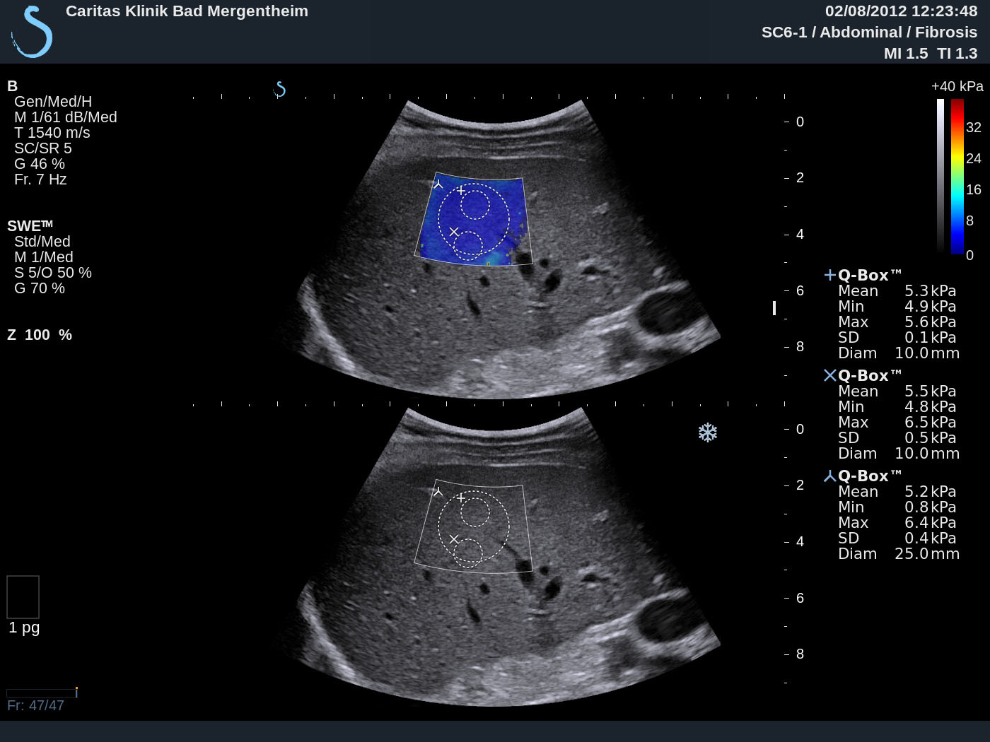
Supersonic Shear Imaging ShearWave Elastography [Dec 2016]
December 11, 2016CEUS LI RADS
January 19, 2017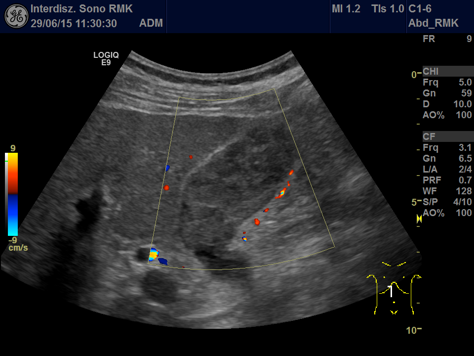
A rare tumor in the gallbladder mimicking sludge
Department for General Internal Medicine and Gastroenterology
Interdisciplinary Sonography Center
Rems-Murr-Klinikum Winnenden gGmbH
Am Jakobsweg 1
D-71364 Winnenden, Germany
Phone: +49 (7195) 591-39322
Mail: Klaus.Dirks@rems-murr-kliniken.de
![A rare tumor in the gallbladder mimicking sludge</br> [Jan 2017]](http://s834315022.websitehome.co.uk/wp-content/uploads/2020/11/cotm_jan2017-fig1.jpg)
![A rare tumor in the gallbladder mimicking sludge</br> [Jan 2017]](http://s834315022.websitehome.co.uk/wp-content/uploads/2020/11/cotm_jan2017-fig2.jpg)
![A rare tumor in the gallbladder mimicking sludge</br> [Jan 2017]](http://s834315022.websitehome.co.uk/wp-content/uploads/2020/11/cotm_jan2017-fig3.jpg)
![A rare tumor in the gallbladder mimicking sludge</br> [Jan 2017]](http://s834315022.websitehome.co.uk/wp-content/uploads/2020/11/cotm_jan2017-fig4.jpg)
![A rare tumor in the gallbladder mimicking sludge</br> [Jan 2017]](http://s834315022.websitehome.co.uk/wp-content/uploads/2020/11/cotm_jan2017-fig5.jpg)
![A rare tumor in the gallbladder mimicking sludge</br> [Jan 2017]](http://s834315022.websitehome.co.uk/wp-content/uploads/2020/11/cotm_jan2017-fig6.jpg)
A 61 year old man was admitted to our hospital due to acute GI-bleeding. The man had been completely healthy until than and did not need any medications. One year ago a small melanoma on the head had been removed in toto. Routine ultrasonography before endoscopy showed echogenic material in the gallbladder (Fig. 1 + 2, Video 1). First gallbladder sludge was suspected. Because of its inhomogeneity in an additional linear array examination and a Doppler examination performed. Here arterial vessels in the gallbladder lumen were detected, thereby proving solid tissue (Video 2).
In addition, a contrast study (CEUS) was performed: This showed multiple polypoid tumors in the gallbladder lumen with strong contrast enhancement in the arterial phase and rapid washout (Fig. 3 + Video 3). Thus the echos in the gallbladder were no sludge but an intraluminal tumor! Only CEUS revealed the extend of tumor masses filling nearly the complete gallbladder.
Unfortunately, the following endoscopy revealed multiple metastases in the stomach, duodenum and colon (Fig. 4-6) that endoscopically and histologically confirmed as metastasis of a malignant melanoma.
Malign gallbladder polyps fortunately are a rare event. Even less common than than gallbladder cancer are metastases within the gallbladder or in the gallbladder wall. Melanoma metastases can be found in nearly every human organ, even in the gallbladder. This case demonstrates melanoma metastases in the gallbladder as well as in the GI tract. Both show a polypoid tumor growth originating from the mucosa.
Echogenic material in the gallbladder lumen mostly is caused by thickened bile: “sludge”. Clumped together sludge may imitate gallbladder polyps. Vice versa polypoid tumors can imitate sludge, especially in cases of hasty US examination. In any case of doubts you should use high-frequency linear probe, examination in left side position und color Doppler. Vessels in the lesion prove a neoplastic polyp witch may be benign or malign.
As shown in this case CEUS is even more sensitive in detecting tumor vascularization. CEUS in case of doubt is an excellent and reliable tool to distinguish between tumor tissue, sludge and concrements.
2) Barretta ML, Catalano O, Setola SV, Granata V, Marone U, D'Errico Gallipoli A. Gallbladder metastasis: spectrum of imaging findings. Abdom Imaging. 2011 Dec;36(6):729-34.
3) Giannini I, Cutrignelli DA, Resta L, Gentile A, Vincenti L. Metastatic melanoma of the gallbladder: report of two cases and a review of the literature. Clin Exp Med. 2015 May 1-6.
Figure 2: Second look using the linear array: Note the inhomogeneity of the echos in the lumen. Left side position does not cause any changes. Also note the well preserved wall of the gallbladder.
Figure 3: CEUS (arterial phase 13 sec after bolus injection): powerful arterial contrast enhancement in multiple polypoid lesions in the lumen of the gallbladder. This proves neoplastic tissue and rules out simple sludge.
Figure 4: Polypoid melanoma metastasis in the duodenum. Note central depression as a sign of malignancy. The neighboring mucosa is completely preserved as well as the neighboring gallbladder wall in figure 1-3.
Figure 5: Polypoid melanoma metastasis in the stomach. Typical black color.
Figure 6. Polypoid melanoma metastasis in the colon. Typical black color.
Video 2: Second look using the linear array and Doppler: Using low PRF several arterial branches inside the gallbladder can be detected. Note the inhomogeneity of the echos in the lumen.
Video 3: Several polyps (= melanoma metastases) within the gallbladder with marked arterial contrast enhancement. Sludge, hematoma or other avital material could definitively be excluded.
