CEUS Liver
December 18, 2015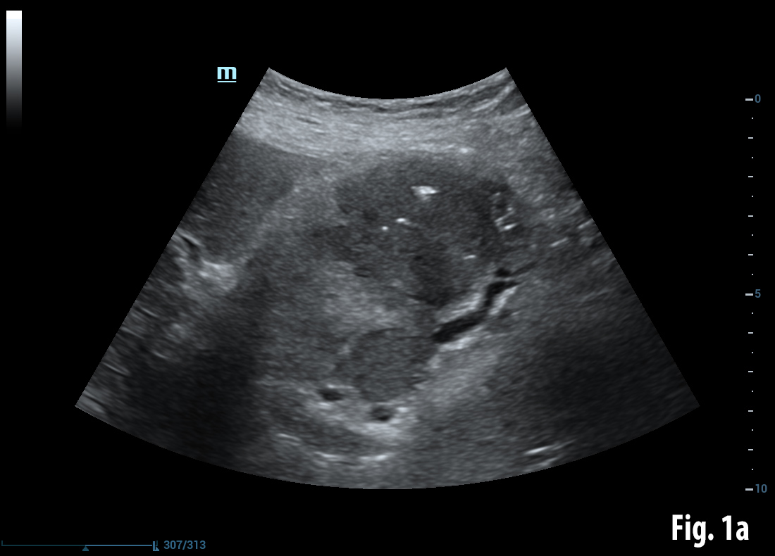
Bouveret syndrome [Apr 2016]
April 10, 2016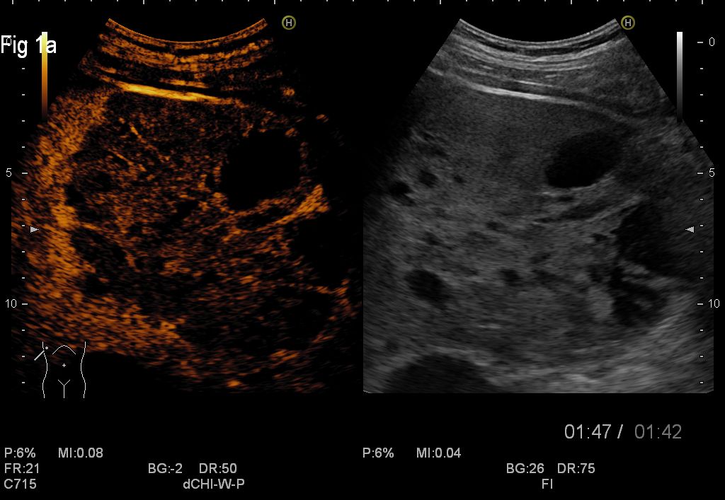
Cystic Liver Metastases of a Neuroendocrine Pancreatic Tumor
Correspondence: Prof. Dr. med. Christoph F. Dietrich
Elena-Codruța Constantinescu1, Larisa Săndulescu1, Carmen Florina Popescu2, Adrian Săftoiu1,3
1. Research Center of Gastroenterology and Hepatology Craiova, University of Medicine and Pharmacy Craiova, Romania
2. Cytology Laboratory, Clinical County Emergency Hospital Craiova, Romania
3. Endoscopy Department, Copenhagen University Hospital Herlev, Denmark
![Cystic Liver Metastases of a Neuroendocrine Pancreatic Tumor</br> [Mar 2016]](http://s834315022.websitehome.co.uk/wp-content/uploads/2020/11/cotm_march2016-fig1a.jpg)
![Cystic Liver Metastases of a Neuroendocrine Pancreatic Tumor</br> [Mar 2016]](http://s834315022.websitehome.co.uk/wp-content/uploads/2020/11/cotm_march2016-fig1b.jpg)
![Cystic Liver Metastases of a Neuroendocrine Pancreatic Tumor</br> [Mar 2016]](http://s834315022.websitehome.co.uk/wp-content/uploads/2020/11/cotm_march2016-fig2.jpg)
1Summary
Pancreatic neuroendocrine tumours (pNET) are considered rare lesions with an incidence of less than 1 per 100 000 person-years. Five-year survival is about 55% when the tumours are localized and resected, but only about 15% when the tumours are not resectable (1). In advanced stages of this disease, as many as 50% of the patients with pNET have already developed metastases in the moment of initial diagnosis, whilst the presence of liver metastases is the major determinant of survival (2). We present a case of a neuroendocrine tumour of the pancreas with liver metastases, imaged with grey-scale, colour Doppler, contrast-enhanced ultrasound (CEUS) and endoscopic ultrasound.
2Discussion
Even though pNETs carry a better prognosis as compared to adenocarcinoma of the pancreas, approximately 90% are silent and non-functional; therefore, most patients are diagnosed in late stage and present metastatic (60%) or locally unresectable advanced disease (21%) with a poor prognosis (3). A specific feature of this case was the presence of cystic metastases of the liver, some with internal septations and some with solid content. Most hepatic metastases are solid, but some have a complete or partially cystic appearance. Hypervascular metastatic tumours with rapid growth may lead to necrosis and cystic degeneration. This mechanism is frequently demonstrated in metastases from neuroendocrine tumours, sarcoma, melanoma, and certain subtypes of lung and breast carcinoma (4).
For this case, CEUS supported the diagnosis of cystic metastases, whilst studies showed previously that CEUS represents a useful method in clinical practice and clearly improves the differential diagnosis between malignant and benign liver lesions detected on standard ultrasonography, with a positive predictive value of 95.4% and negative predictive value of 95.9%. The main criteria for malignancy on CEUS is contrast wash-out in the late portal venous phase whereas benign lessons typically remain iso-enhancing with the surrounding normal liver tissue (5,6). Metastases usually show a brief arterial hypervascularity and complete rapid wash-out, which can improve detection during the portal phase (7). Neoplastic cysts such as cystic metastasis can be characterized on CEUS by sensitive real-time demonstration of vascular flow within the septa or solid component. Non-neoplastic complex cysts such as hemorrhagic cysts or hydatid cysts show the absence of intralesional enhancement on CEUS, thus confirming their non-neoplastic nature (8).
For this case, CEUS supported the diagnosis of cystic metastases, whilst studies showed previously that CEUS represents a useful method in clinical practice and clearly improves the differential diagnosis between malignant and benign liver lesions detected on standard ultrasonography, with a positive predictive value of 95.4% and negative predictive value of 95.9%. The main criteria for malignancy on CEUS is contrast wash-out in the late portal venous phase whereas benign lessons typically remain iso-enhancing with the surrounding normal liver tissue (5,6). Metastases usually show a brief arterial hypervascularity and complete rapid wash-out, which can improve detection during the portal phase (7). Neoplastic cysts such as cystic metastasis can be characterized on CEUS by sensitive real-time demonstration of vascular flow within the septa or solid component. Non-neoplastic complex cysts such as hemorrhagic cysts or hydatid cysts show the absence of intralesional enhancement on CEUS, thus confirming their non-neoplastic nature (8).
3References
1) Exocrine and endocrine pancreas. In: Edge SB, Byrd DR, Compton CC, et al., eds.: AJCC Cancer Staging Manual. 7th ed. New York, NY: Springer, 2010, pp 241-9.
2) Pattou F, Proye C. Endocrine tumors of the pancreas. In: Holzheimer RG, Mannick JA, editors. Surgical Treatment: Evidence-Based and Problem-Oriented. Munich: Zuckschwerdt; 2001
3) Orditura M, Petrillo A, Ventriglia J, Diana A, Laterza MM, Fabozzi A, Savastano B, Franzese E, Conzo G, Santini L, Ciardiello F, De Vita F. Pancreatic neuroendocrine tumors: nosography, management and treatment (review). 2015 Dec 17. S1743-9191(15)01438-7
4) Lewis KH, Chezmar JL. Hepatic metastases. Magn Reson Imaging Clin N Am 1997;5:319-330.
5) Strobel D, Seitz K, Blank W, Schuler A, Dietrich C, von Herbay A, Friedrich-Rust M, Kunze G, Becker D, Will U, Kratzer W, Albert FW, Pachmann C, Dirks K, Strunk H, Greis C, Bernatik T. Contrast-enhanced ultrasound for the characterization of focal liver lesions--diagnostic accuracy in clinical practice (DEGUM multicenter trial). Ultraschall Med. 2008;29:499-505.
6) Sporea I, Badea R, Popescu A, Spârchez Z, Sirli RL, Dănilă M, Săndulescu L, Bota S, Calescu DP, Nedelcu D, Brisc C, Ciobâca L, Gheorghe L, Socaciu M, Martie A, Ioaniţescu S, Tamas A, Streba CT, Iordache M, Simionov I, Jinga M, Anghel A, Cijevschi Prelipcean C, Mihai C, Stanciu SM, Stoicescu D, Dumitru E, Pietrareanu C, Bartos D, Manzat Saplacan R, Pârvulescu I, Vădan R, Smira G, Tuţă L, Săftoiu A. Contrast-enhanced ultrasound (CEUS) for the evaluation of focal liver lesions - a prospective multicenter study of its usefulness in clinical practice. Ultraschall Med. 2014 Jun;35(3):259-66. doi: 10.1055/s-0033-1355728. Epub 2014 Feb 21. PMID: 24563420
7) Hyun-Jung Jang, Hojun Yu, Tae Kyoung Kim. Contrast-enhanced ultrasound in the detection and characterization of liver tumors. 2009 Nov 6. doi: 10.1102/1470-7330.2009.0015
8) Kim TK, Jang HJ, Wilson SR. Benign liver masses: imaging with microbubble contrast agents.Ultrasound Q. 2006;22:31–9
4Figures
Figure 1a,1b. CEUS of the secondary cystic liver masses in the late venous phase
Figure 2. EUS demonstrates the primary tumour in the pancreas as a heterogenous hypo-and hyperechoic mass
Movie 1. Contrast-enhanced ultrasound of the cystic metastases during the arterial and portal venous phases.
Movie 2. Contrast-enhanced EUS showing the malignant pNET in the head of the pancreas during the early arterial and late venous phases
