TITLE: Acute appendicitis
DESCRIPTION:
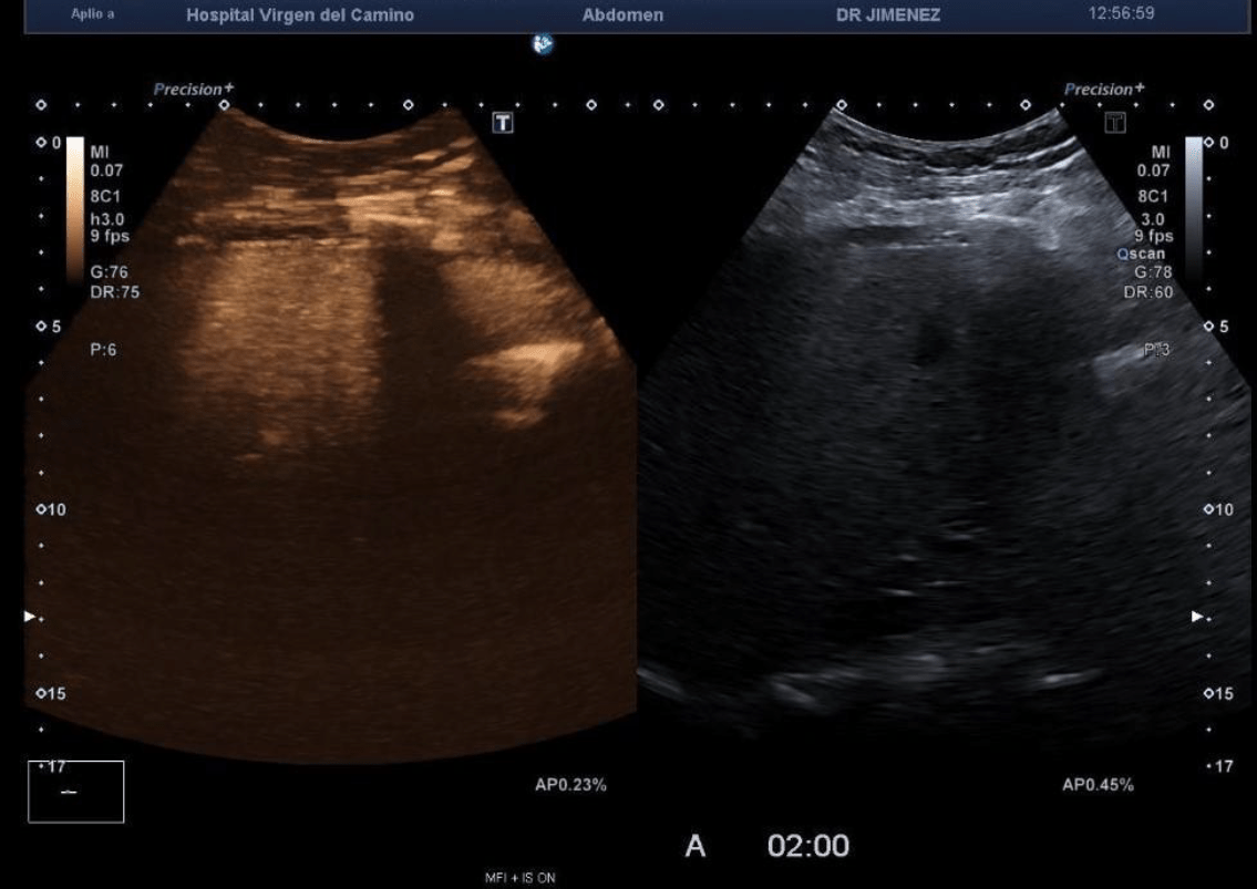
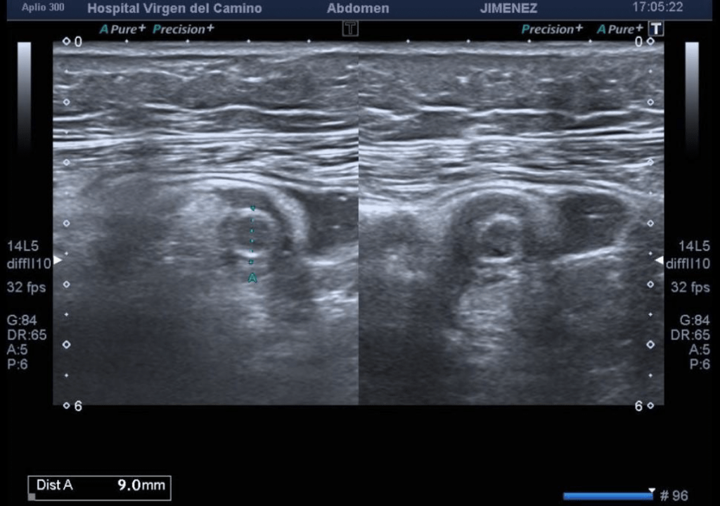
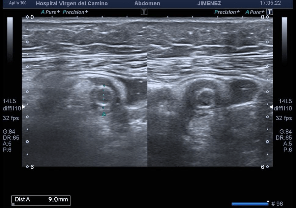
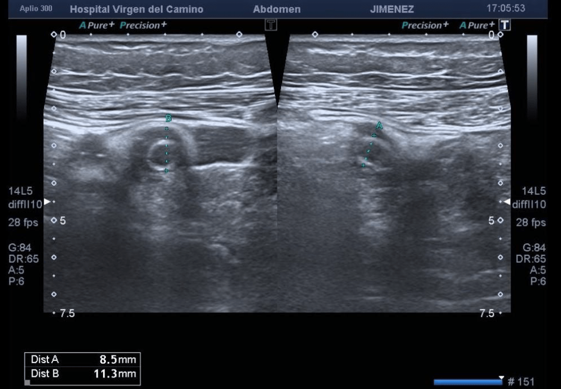
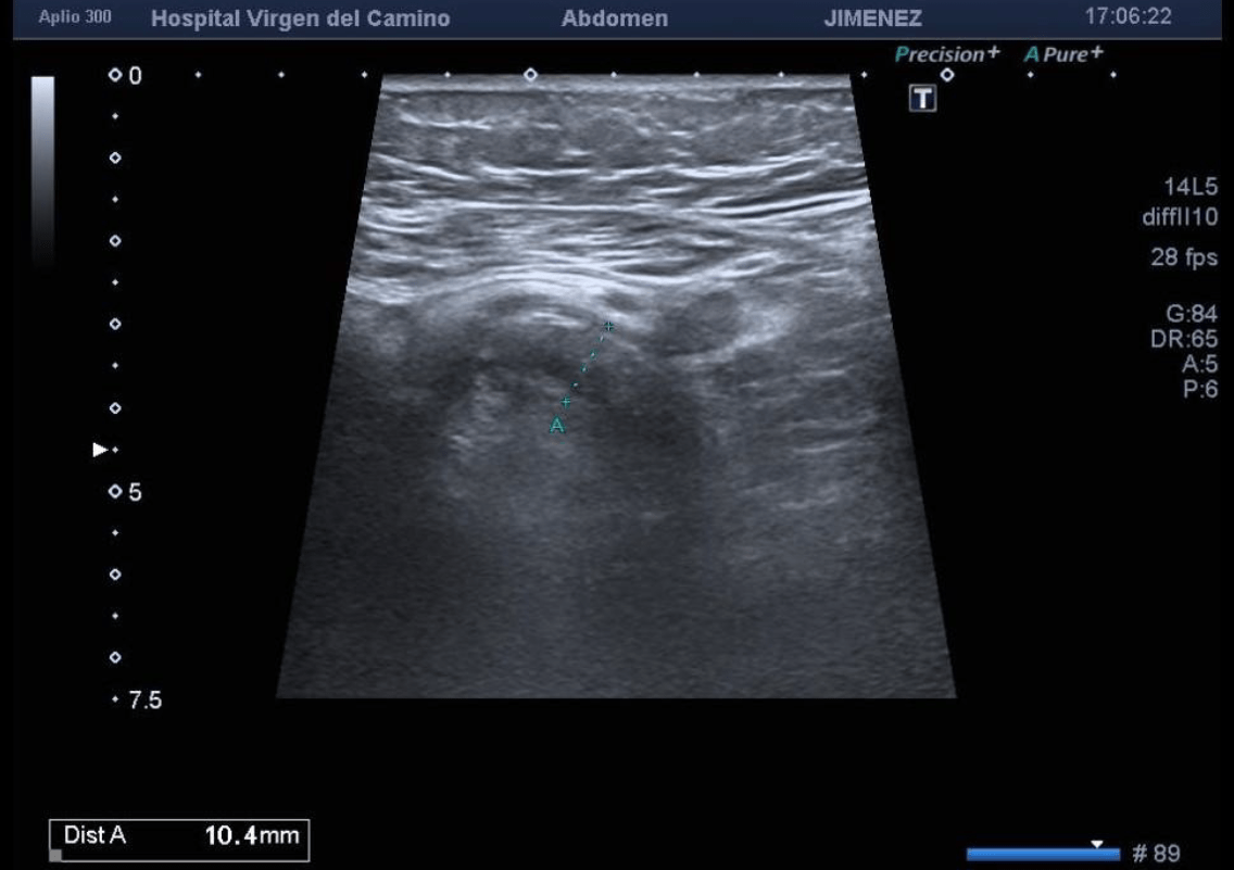
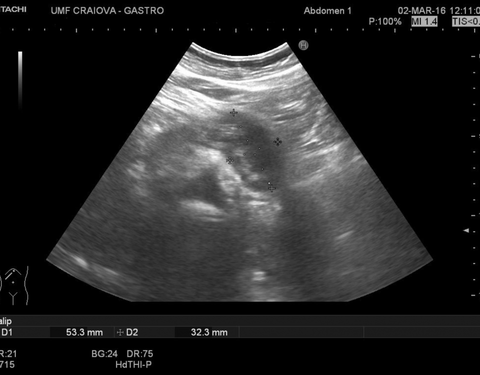
a 63 years old female was seen in the emergency department with pain in the right iliac fossa. By ultrasound we identify a non-peristaltic and non-compressible tubular structure with external compression maneuvers in the right iliac fossa. It originates at the base of the cecum and terminates in the cul-de-sac, consistent with the cecal appendix. It has has a diameter of 10 mm and is accompanied by a small amount of free fluid in the pelvis as well as inflammatory changes in the adjacent fat. Positive ultrasound rebound tenderness
(Blumberg sign).
AUTHORS: Miguel Emilio Oliver Pece y Sebastián Jiménez Sánchez. Hospital Virgen del Camino, Sanlúcar de Barrameda, Cádiz, España.
EMAIL CONTACT:
moliverp@hotmail.com
KEYWORDS:
acute apendicitis