
TITLE: Hemangioma 004
DESCRIPTION:
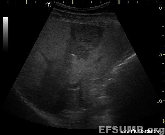
Hyperechoic liver lesion inside a hypoechoicarea (Figure 1, Movie 1) in a patient with non-alcoholic liver steatosis and overweight.
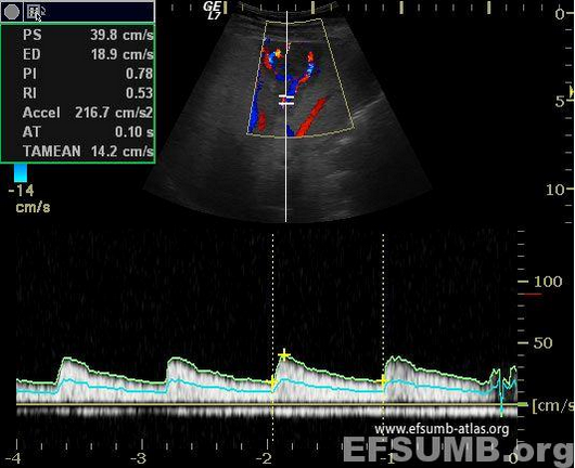
Doppler Color examination reveals the feeding artery (Figure 2, Movie 2).
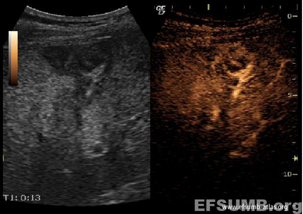
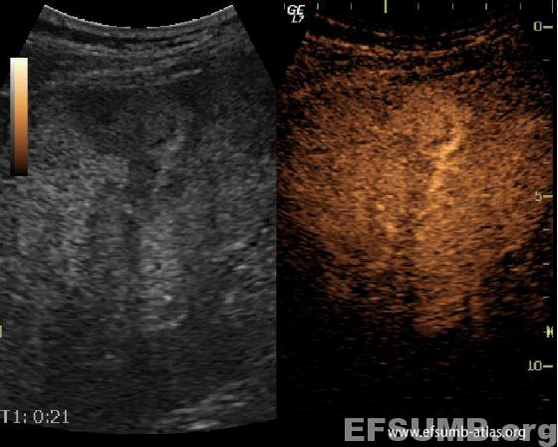
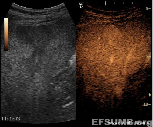
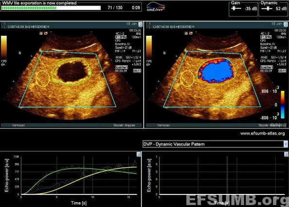
The structural change was followed after intravenous contrast agent administration (SonoVue). During the arterial phase it’s shown the contrast agent enhancement of the nodule in a centripetal manner, by an intake vessel (Figure 3, Figure 4, Movie 3). No wash-out was seen during the portal and venous phases (Figure 5, Figure 6, Movie 4). The surrounding hypoechoic area presents homogenous enhancement but slightly delayed compared to the surrounding parenchyma.
The diagnosis was of an arterialized hemangioma in a fatty liver, with vascular changes trough vascular steal phenomenon.
[Sirli R et al, Med Ultrason 2011; 13(2):95-101]
[Wilson SR et al, J Ultrasound Med 2007; 26: 775-787]
Courtesy of:
BadeaRadu (1), LilianaChiorean (2)(*)
1) Department of Ultrasonography, “Octavian Fodor” Institute of Gastroenterology and Hepatology, “IuliuHatieganu” University of Medicine and Pharmacy, Cluj-Napoca, Romania
2) Department of Radiology and Computed Tomography, “Octavian Fodor” Institute of Gastroenterology and Hepatology, Cluj-Napoca, Romania
(*) Corresponding author:
LilianaChiorean
Department of Radiology and Computed Tomography, “Octavian Fodor” Institute of Gastroenterology and Hepatology Str. Croitorilor 19 – 21, 400 162 ClujNapoca, Romania
KEYWORDS:
hemangioma, non-cirrhotic liver, focal liver lesions, CEUS