Incorrect ....Please see the correct answer highlighted
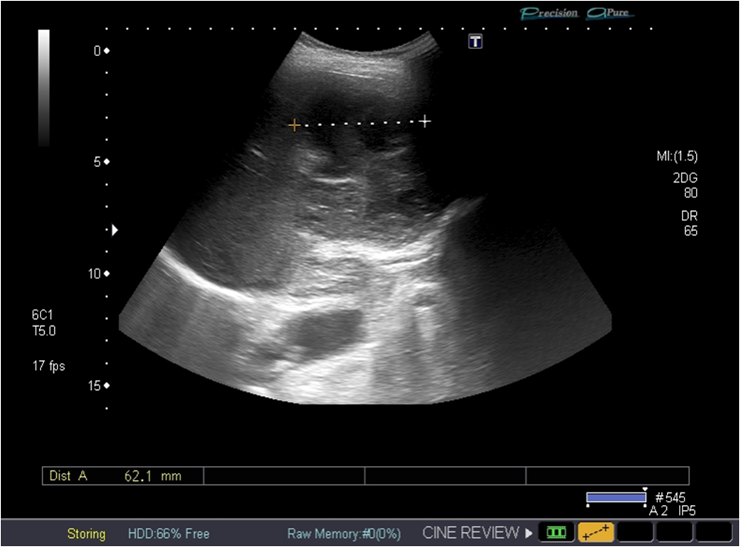
Ultrasound showed splenomegaly and a solid mass tumor: a large hypoechoic focal lesion located near the splenic hillum Further assesment with contrast enhanced ultrasound revealed a rapid influx of the contrast agent, resulting in enhancement in the arterial phase, non-enhancing in the middle and intense wash-out in late phases.
Splenectomy was performed and histopathological examination confirmed the diagnosis of splenic Burkitt lymphoma.

