Incorrect ....Please see the correct answer highlighted
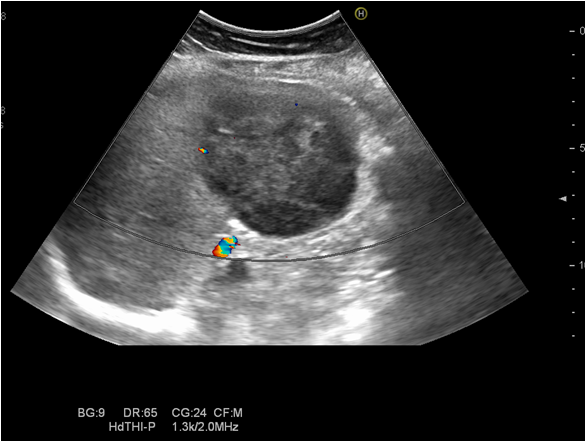
Standard ultrasound showed two oval lesions in the right liver lobe, segment VIII, poorly demarcated, with a mixt complex pattern, a transverse diameter of 7 and 4 cm and another 3,5 cm hypoechoic lesion situated in segment VI. Image guided drainage was performed, with a favourable evolution.
Standard ultrasound showed two oval lesions in the right liver lobe, segment VIII, poorly demarcated, with a mixt complex pattern, a transverse diameter of 7 and 4 cm and another 3,5 cm hypoechoic lesion situated in segment VI. Image guided drainage was performed, with a favourable evolution.

