Student Image Challenge #92
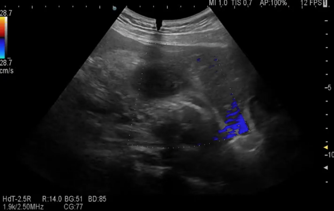
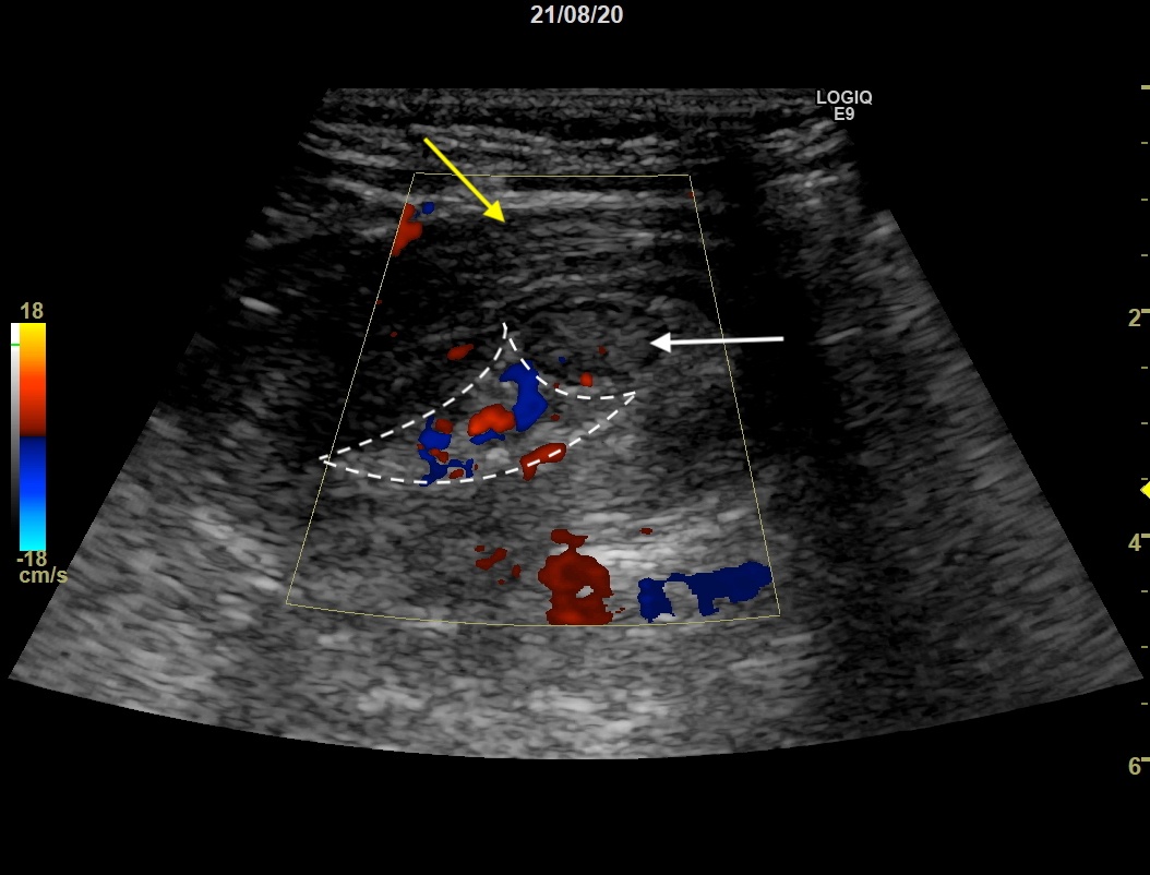
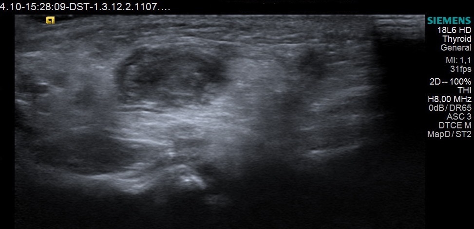
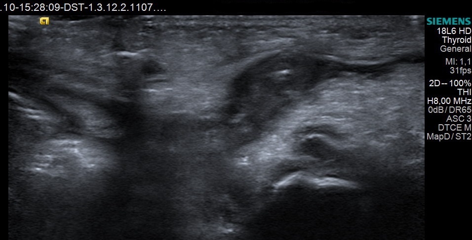
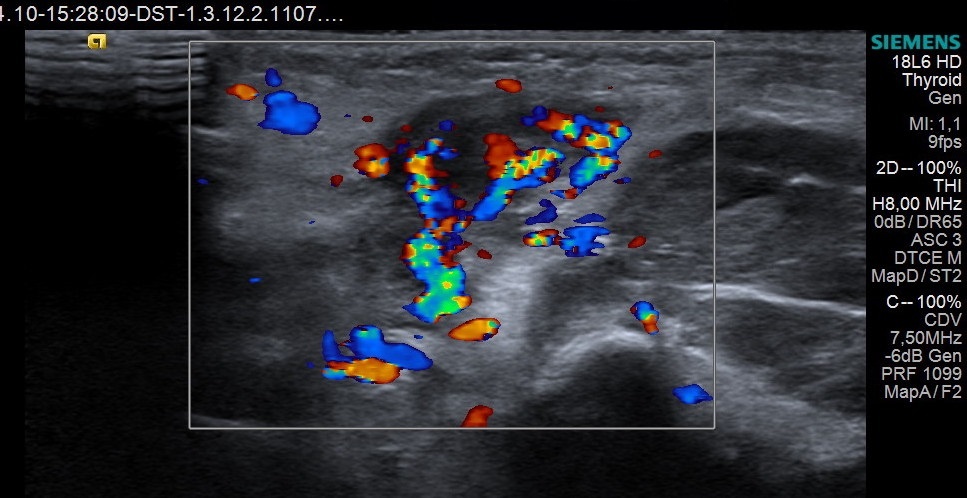
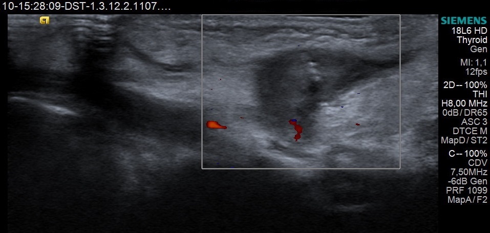
A 2-month-old girl was surgically treated for a large congenital inguinal hernia, which also included the fallopian tube. The postoperative evaluation revealed in the groin area a tubular parenchymal structure coming from the abdominal cavity, with glove-finger-like ending. Moreover, distally to the respective structure, there were some subtile tubular structures with a small amount of fluid.
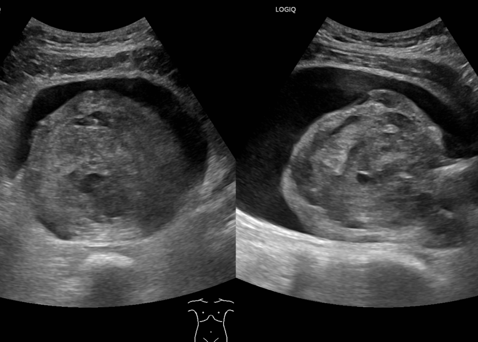
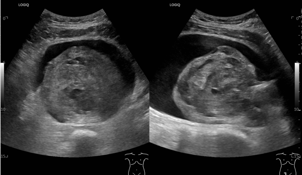
The pelvic examination showed intestinal loops with discrete stasis, between which the ovary could not be detected with certainty. Given the imaging uncertainty, it was re-intervened and the inguinal orifice was considered normal.
After 2 days, the pathological structure decreased in size and especially in terms of the number of vessels in Color Doppler examination.
What would be your diagnosis?
Incorrect ....Please see the correct answer highlighted