TITLE: Hemangioma 001
DESCRIPTION:
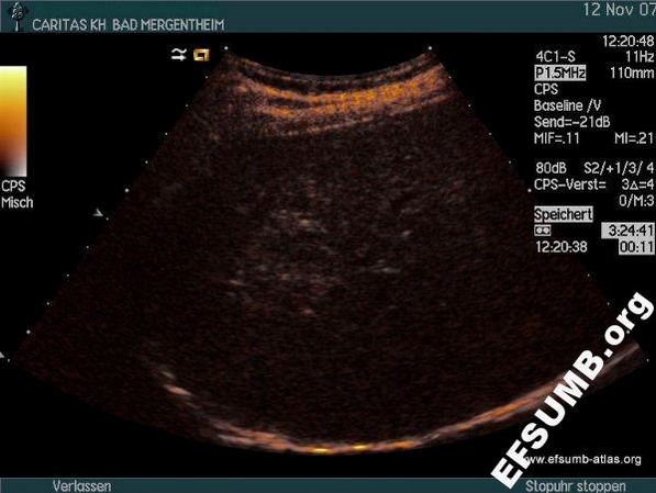
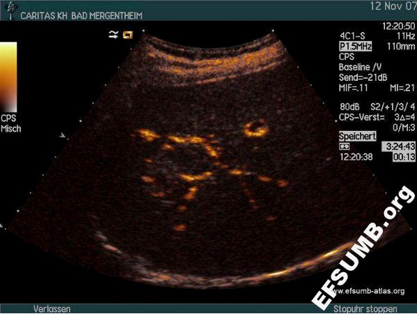
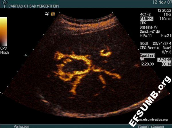
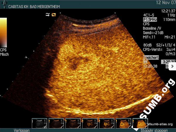
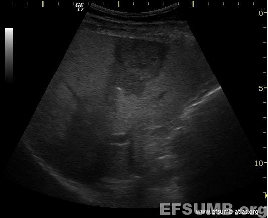
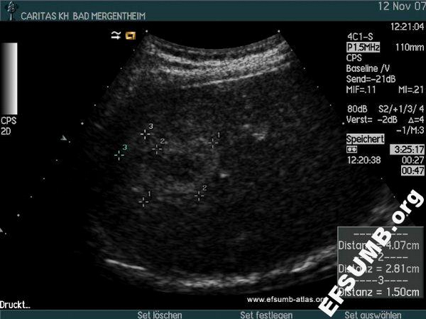
Hemangioma with typical peripheral nodular contrast enhancement and centripetal fill-in. The lesion is displayed in the conventional B-mode scanning (fig.1) and in contrast images (fig. 2 - 7) [EFSUMB Case of the Month, www.EFSUMB.org].
KEYWORDS:
hemangioma peripheral nodular contrast enhancement centrepetal lesion b-mode scanning