
TITLE: Hemangioma 002
DESCRIPTION:
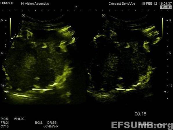
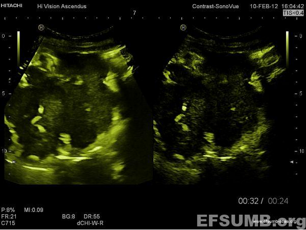
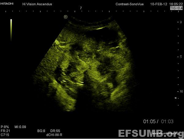
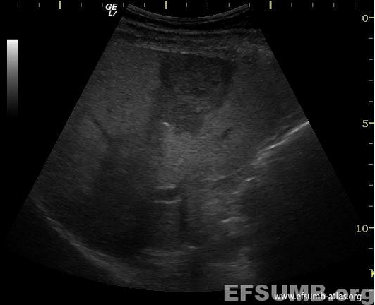
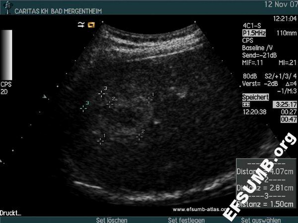
Hemangioma (so-called giant hemangioma [{Dietrich, 2007 2945 /id}]).
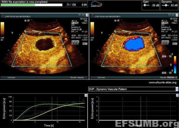
B-mode (1), arterial (2,3) (using Microbubble Tracing Imaging [MTI]) and portal venous phases (4) are shown with periperal nodular contrast enhancement and centripetal fill in. It is important for diagnosis that the nodules are hyperenhancing during all phases and that bubble destruction is avoided [Dietrich CF, Maddalena ME, Cui XW, Schreiber-Dietrich D, Ignee A. Liver tumor characterization – review of the literature. Ultraschall (Suppl) 2012;33:S3-S10].
KEYWORDS:
hemangioma b-mode arterial microbubble tracing imaging portal venous phases peripheral nodular contrast enhancement