Ultrasound diagnosis of uncomplicated interstitial pregnancy [May 2016]
May 10, 2016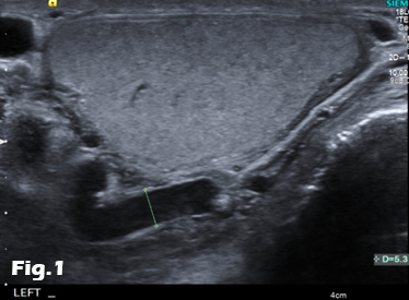
Scrotal varicocele in older patients: should we search for a renal tumour? [Jul 2016]
July 11, 2016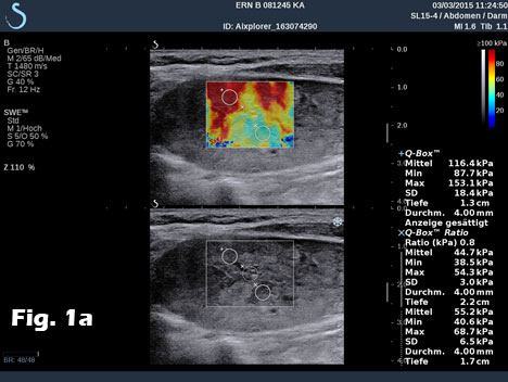
Schwannoma and jugular vein thrombosis diagnosed by using modern imaging technology
Prof. Dr. med. Christoph F. Dietrich
Medizinische Klinik 2, Caritas-Krankenhaus, Uhlandstr. 7
97980 Bad Mergentheim Tel:+49 7931 58 2201
Email: christoph.dietrich@ckbm.de
Medizinische Klinik 2, Caritas-Krankenhaus, Uhlandstr. 7
97980 Bad Mergentheim Tel:+49 7931 58 2201
Email: christoph.dietrich@ckbm.de
![Schwannoma and jugular vein thrombosis diagnosed by using modern imaging technology</br> [Jun 2016]](http://s834315022.websitehome.co.uk/wp-content/uploads/2020/11/cotm-june2016-fig1a.jpg)
![Schwannoma and jugular vein thrombosis diagnosed by using modern imaging technology</br> [Jun 2016]](http://s834315022.websitehome.co.uk/wp-content/uploads/2020/11/cotm-june2016-fig1b.jpg)
![Schwannoma and jugular vein thrombosis diagnosed by using modern imaging technology</br> [Jun 2016]](http://s834315022.websitehome.co.uk/wp-content/uploads/2020/11/cotm-june2016-fig1c.jpg)
![Schwannoma and jugular vein thrombosis diagnosed by using modern imaging technology</br> [Jun 2016]](http://s834315022.websitehome.co.uk/wp-content/uploads/2020/11/cotm-june2016-fig1d.jpg)
![Schwannoma and jugular vein thrombosis diagnosed by using modern imaging technology</br> [Jun 2016]](http://s834315022.websitehome.co.uk/wp-content/uploads/2020/11/cotm-june2016-fig1e.jpg)
1Summary
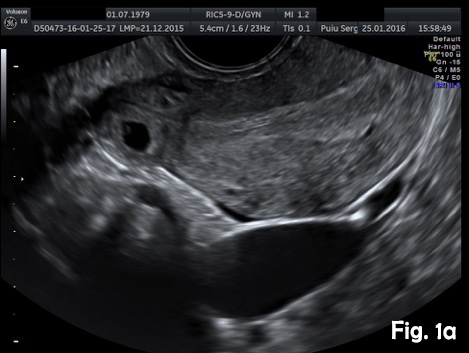
We report on the incidental finding of a 70 y/o patient presenting with an indolent cervical swelling and mass. Clinical examination and laboratory data were not conclusive. In the figure sequence B mode ultrasound and shear wave elastography revealed a 43 mm mass lesion with peripherally stiffer tissue (a) with thrombosis in statu nascendi of jugular vein shown by B mode (b) and also by contrast enhanced ultrasound (c) and shear wave elastography (d). 3D shear wave elastography confirmed the circumscript peripherally stiffer cervical mass (e). 3D SWE added complementary information for a better delineation of the distribution of increased stiffness, following the fibrous capsule around the nerve. SWE also showed feasibility to demonstrate stiffer tissue within jugular vein. 3D techniques allowed improved documentation of the anatomical volume including the tumour and surrounding vessels [(1-3)]. Histologically the nerve tumour schwannoma was proven.
2Discussion
Schwannoma (synonymous "Schwann cell tumor", neurolemoma or neurilemoma) is most often slowly growing benign tumour deriving from Schwann cells (S-100 positive) of central or peripheral located nerves. It is the most common tumour of peripheral nerves. Only few schwannoma (less than 10 %) are associated with neurofibromatosis type 1. Malignant transformation is rare (neurofibrosarcoma). Most often they are found incidentally.
Symptoms are caused by expansion into the surrounding structures. The treatment options include observation in carefully selected patients, surgery or radiation therapy depending on their location, symptoms, age of the patient and comorbidity. Schwannomas show a biphasic architecture of Antoni A (dense) and B (loose) patterns, as well as nuclear palisading (Verocay bodies) and a fibrous capsule containing the parent nerve [(4)]. Differential diagnosis include inflammatory and neoplastic lymphadenopathy and other primary or secondary tumours [(5-12)].
3Shear wave elastography (SWE)
Ultrasound-based shear wave elastography is an imaging mode that enables an ultrasound platform to quantify tissue elasticity with or without an elasticity map, depending on its technical implementation. In all commercially available ultrasound systems, it uses the acoustic radiation force related to ultrasound focusing to generate shear waves in tissue. These mechanical shear waves then propagate orthogonally to the pushing ultrasound beam at a speed that can be assumed to be proportional to the square root of tissue Young’s Modulus, also known as tissue elasticity [(13;14)]. Here we used ShearWave™ Elastography (SWE™) that was implemented by SuperSonic Imagine on the Aixplorer® ultrasound imaging system. This ultrasound platform is capable of reaching an acquisition frame rate that is suitable with the supersonic shear imaging (SSI) technique [(15)]. This technical implementation of shear wave elastography enables the operator to get a quantitative mapping of tissue elasticity in real-time, at an imaging frame rate of a few images per second. Thanks to the supersonic shear imaging technique and the ultrafast acquisition speed (several thousands of images per second), heating of the probe is avoided and acoustic energy stays within the limits used in routine so no cool-down time is required, operators are getting a real-time feedback like with any other ultrasound imaging mode, thus allowing them to adjust their scanning technique as required, and many trade-offs can be saved between the overall image quality, the size and depth of the SWE-Box, the spatial and temporal resolutions in SWE™, and the maximum elasticity values measured.
43D SWE
On Aixplorer®, ShearWave™ Elastography has also been implemented on a linear 3D wobbling probe. Leveraging on the ultrafast imaging capabilities of Aixplorer, the acquisition of both grayscale imaging and SWE™ imaging in a three-dimensional volume takes a few seconds, depending on the size of the volume, and data can be analysed and reprocessed retrospectively in the full volume. Thanks to the high frequency broad bandwidth transducer (5 to 16 MHz), high quality images can be reconstructed in the axial, the transverse and the coronal planes. The operator can choose to navigate in the volume acquired using a customizable multislice or multiplanar display. The probe was mainly designed to be used in breast imaging. Here we reported on the first use of 3D SWE in nerve tumours. 3D SWE added complementary information for a better delineation of the stiffer fibrous capsule around the nerve.
5Review of the literature (3D SWE)
In the field of breast lesions and breast cancer imaging, 3D SWE™ has been reported to provide equivalent results as compared to 2D SWE™ for the characterization of known breast masses [(16)]. Using SWE™ to help in characterizing 144 breast masses with ultrasound, Lee et al reported an increase in specificity of breast ultrasound from 30% up to 64% with 3D SWE, without any significant change in sensitivity. In another study on 146 patients with 163 breast masses, it was demonstrated that the inter-observer agreement on breast cancer risk assessment with ultrasound was significantly increased from kappa=0.38 to kappa=0.73 with the addition of 3D-SWE qualitative assessment [(17)]. Still in the breast arena, Athanasiou et al reported recently that tumor volume assessment with 3D ultrasound and 3D SWE was highly concordant with dynamic contrast-enhanced MRI tumour volume. In this feasibility study on 10 patients, 3D SWE demonstrated a clear value as a potential indicator of breast cancer response to neoadjuvant chemotherapy, because it could assess at the same time the changes in tumour volume and stiffness [(18)].
6References
1. Dietrich CF. 3D abdominal sonography. Electromedica 2001; 69(2):23-29.
2. Dietrich CF. [3D real time contrast enhanced ultrasonography,a new technique]. Rofo 2002; 174(2):160-163.
3. Hocke M, Dietrich CF. New technology--combined use of 3D contrast enhanced endoscopic ultrasound techniques. Ultraschall Med 2011; 32(3):317-318.
4. Skovronsky DM, Oberholtzer JC. Pathologic classification of peripheral nerve tumors. Neurosurg Clin N Am 2004; 15(2):157-166.
5. Chiorean L, Barr RG, Braden B, Jenssen C, Cui XW, Hocke M et al. Transcutaneous Ultrasound: Elastographic Lymph Node Evaluation. Current Clinical Applications and Literature Review. Ultrasound Med Biol 2016; 42(1):16-30.
6. Cui XW, Jenssen C, Saftoiu A, Ignee A, Dietrich CF. New ultrasound techniques for lymph node evaluation. World J Gastroenterol 2013; 19(30):4850-4860.
7. Cui XW, Ignee A, Bachmann NM, Schreiber-Dietrich D, De Molo C, Pirri C et al. Contrast enhanced ultrasound of sentinel lymph nodes. J Ultrason 2013; 13:73-81.
8. Cui XW, Hocke M, Jenssen C, Ignee A, Klein S, Schreiber-Dietrich D et al. Conventional ultrasound for lymph node evaluation, update 2013. Z Gastroenterol 2014; 52(2):212-221.
9. Cui XW, Chang JM, Kan QC, Chiorean L, Ignee A, Dietrich CF. Endoscopic ultrasound elastography: Current status and future perspectives. World J Gastroenterol 2015; 21(47):13212-13224.
10. Dietrich CF, Annema JT, Clementsen P, Cui XW, Borst MM, Jenssen C. Ultrasound techniques in the evaluation of the mediastinum, part I: endoscopic ultrasound (EUS), endobronchial ultrasound (EBUS) and transcutaneous mediastinal ultrasound (TMUS), introduction into ultrasound techniques. J Thorac Dis 2015; 7(9):E311-E325.
11. Dietrich CF, Jenssen C, Arcidiacono PG, Cui XW, Giovannini M, Hocke M et al. Endoscopic ultrasound: Elastographic lymph node evaluation. Endosc Ultrasound 2015; 4(3):176-190.
12. Dietrich CF, Annema JT, Clementsen P, Cui XW, Borst MM, Jenssen C. Ultrasound techniques in the evaluation of the mediastinum, part I: endoscopic ultrasound (EUS), endobronchial ultrasound (EBUS) and transcutaneous mediastinal ultrasound (TMUS), introduction into ultrasound techniques. J Thorac Dis 2015; 7(9):E311-E325.
13. Bamber J, Cosgrove D, Dietrich CF, Fromageau J, Bojunga J, Calliada F et al. EFSUMB guidelines and recommendations on the clinical use of ultrasound elastography. Part 1: Basic principles and technology. Ultraschall Med 2013; 34(2):169-184.
14. Cosgrove D, Piscaglia F, Bamber J, Bojunga J, Correas JM, Gilja OH et al. EFSUMB Guidelines and Recommendations on the Clinical Use of Ultrasound Elastography.Part 2: Clinical Applications. Ultraschall Med 2013; 34(3):238-253.
15. Bercoff J, Tanter M, Fink M. Supersonic shear imaging: a new technique for soft tissue elasticity mapping. IEEE Trans Ultrason Ferroelectr Freq Control 2004; 51(4):396-409.
16. Lee SH, Chang JM, Kim WH, Bae MS, Cho N, Yi A et al. Differentiation of benign from malignant solid breast masses: comparison of two-dimensional and three-dimensional shear-wave elastography. Eur Radiol 2013; 23(4):1015-1026.
17. Youk JH, Gweon HM, Son EJ, Chung J, Kim JA, Kim EK. Three-dimensional shear-wave elastography for differentiating benign and malignant breast lesions: comparison with two-dimensional shear-wave elastography. Eur Radiol 2013; 23(6):1519-1527.
18. Athanasiou A, Latorre-Ossa H, Criton A, Tardivon A, Gennisson JL, Tanter M. Feasibility of Imaging and Treatment Monitoring of Breast Lesions with Three-Dimensional Shear Wave Elastography. Ultraschall Med 2015.
7Figures
Figure 1:B mode ultrasound and shear wave elastography revealed a 43 mm mass lesion with peripherally stiffer tissue (a) with thrombosis in statu nascendi of jugular vein (b), also shown by contrast enhanced ultrasound (c) and shear wave elastography (d). 3D shear wave elastography confirmed the circumscript peripherally stiffer cervical mass (e).
