
Typical ultrasound features in open spina bifida [January 2022]
January 26, 2022
Neonatal Cranial Ultrasound – Safety Aspects [Update 2022]
March 12, 2022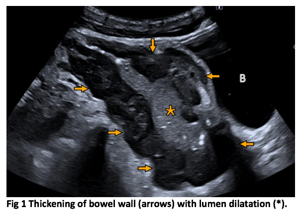
Small intestine lymphoma with aneurysmal dilatation
AUTHOR
Rubén Ruiz Marco
Tomás Ripollés González
María J Martínez-Pérez
Department of Radiology, Dr Peset Hospital, Valencia, Spain
Rubén Ruiz Marco
Tomás Ripollés González
María J Martínez-Pérez
Department of Radiology, Dr Peset Hospital, Valencia, Spain
1Clinical History
A 28-year-old man referred from the emergency department due to diffuse abdominal pain of several days of evolution. Mild weight loss in the last few months. Ultrasound was requested to rule out appendicitis.
2Image findings
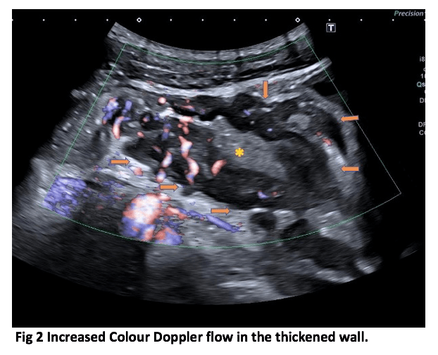
Sagittal US scan of the pelvis showed marked hypoechoic thickening of the wall of an intestinal segment of 11 cm (between arrows), with loss of the wall ecostructure and widening of the echogenic lumen (*) (figure 1). There was slight dilatation of the proximal loops (shown in the video 1). No thickening of the walls of other intestinal segments were detected. B = bladder. Color Doppler showed increased vascularization suggesting mural hyperaemia (figure 2).
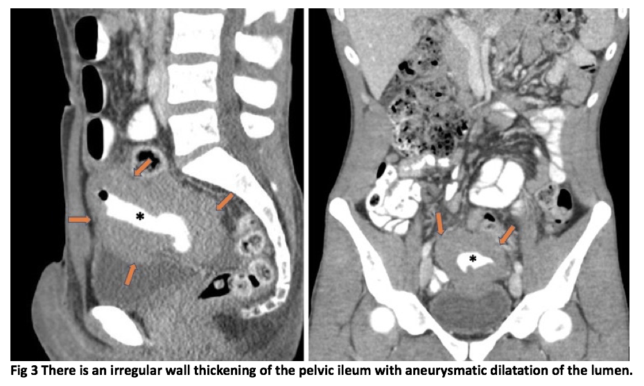
CT performed after IV and oral radiopaque contrast shows a pelvic mass that originates in the wall of the middle ileum, affecting a segment of 12 cm, with mural thickening. Passage of radiopaque oral contrast through its dilated lumen is observed, without extraluminal extravasation or associated abscess (Figure 3). There is dilatation of proximal intestinal loops (Figure 4).
CT performed after IV and oral radiopaque contrast shows a pelvic mass that originates in the wall of the middle ileum, affecting a segment of 12 cm, with mural thickening. Passage of radiopaque oral contrast through its dilated lumen is observed, without extraluminal extravasation or associated abscess (Figure 3). There is dilatation of proximal intestinal loops (Figure 4).
3Diagnosis
Diffuse large B cell lymphoma of small intestine with aneurysmatic dilatation.
4Discussion
BLymphomas make up about 20% of all small bowel tumours. Approximately 30% of gastrointestinal lymphomas occur in the small intestine. These lymphomas may be broadly categorized into three main groups: Immunoproliferative small intestinal disease lymphoma (IPSID), enteropathy-associated T cell lymphoma and other western-type non-IPSID lymphomas, like the diffuse large B cell lymphoma of this case.
The distal ileum is the most common site, owing to the large amount of lymphoid tissue that is present in the distal ileum. Risk factors include celiac disease, Crohn´s disease, Systemic Lupus Erythematosus, immunocompromised state and a history of chemotherapy or extra-intestinal lymphoma.
Clinical presentation of patients with lymphoma of the small intestine differs according to the histologic tumour type. Patients with IPSID typically present with abdominal pain, chronic diarrhoea, malabsorption, severe weight loss, clubbing, and ankle edema.
Patients with other non-IPSID lymphomas have a more nonspecific presentation, which may include abdominal pain, GI bleeding, intestinal obstruction or perforation, obstructive jaundice, and/or a palpable abdominal mass.
The typical cross-sectional image (US or CT) presentation of a small bowel lymphoma is a thick-walled infiltrating mass with aneurysmal dilatation without obstruction. Aneurysmal dilatation is based upon destruction of the bowel wall and the myenteric nerve plexus.
A less common presentation is as an intraluminal polypoid mass or a large eccentric mass with extension into the surrounding soft tissues with possible ulceration and formation of fistulas.
Large adenocarcinomas and lymphomas can have similar imaging appearances. Bulky mesenteric or retroperitoneal lymphadenopathy and splenomegaly are findings that support the diagnosis of a lymphoma, while infiltration of the mesenteric fat and bowel obstruction favours the diagnosis of an adenocarcinoma.
5Teaching Points
Intestinal lymphoma is a relatively infrequent entity that can be found in ultrasound studies. The main diagnostic challenge using US it is to be able to define the margins of the lesion and verify its dependence of the bowel loops. CT study with oral contrast helps to confirm the relationship of the lesion with the intestinal loops. Once this point is clear, the main differential diagnosis of irregular intestinal thickening is an infectious process (such as tuberculosis) or different tumour aetiologies (mainly adenocarcinoma).
6References
1. Hasaballah M, Abdel-Malek R, Zakaria Z, Marie MS, Naguib MS. Transabdominal ultrasonographic features in the diagnosis of gastrointestinal lymphoma. J Gastrointest Oncol. 2018;9(6):1190-1197. doi:10.21037/jgo.2018.09.02
2. Roccarina D, Garcovich M, Ainora ME, et al. Diagnosis of bowel diseases: The role of imaging and ultrasonography. World J Gastroenterol 2013;19:2144-53. 10.3748/wjg.v19.i14.214
3. Koch P, del Valle F, Berdel WE, et al. Primary gastrointestinal non-Hodgkin's lymphoma: I. Anatomic and histologic distribution, clinical features, and survival data of 371 patients registered in the German Multicenter Study GIT NHL 01/92. J Clin Oncol 2001;19:3861-73. 10.1200/JCO.2001.19.18
4. Cui, Ning-Yi & Gong, Xuan-Tong & Tian, Yan-Tao & Wáng, Yì-Xiáng & Zhang, Rui & Liu, Meng-Jia & Han, Jie & Wang, Bo & Yang, Di. (2021). Contrast-enhanced ultrasound imaging for intestinal lymphoma. World Journal of Gastroenterology. 27. 5438-5447. 10.3748/wjg.v27.i32.5438..3861
